J Vet Sci.
2014 Jun;15(2):195-198. 10.4142/jvs.2014.15.2.195.
Accuracy of sonographic diagnosis of pneumoperitoneum using the enhanced peritoneal stripe sign in beagle dogs
- Affiliations
-
- 1Research Institute of Life Sciences, Gyeongsang National University, Jinju 660-701, Korea. lhc@gnu.ac.kr
- KMID: 1784639
- DOI: http://doi.org/10.4142/jvs.2014.15.2.195
Abstract
- The objective of this study was to evaluate the feasibility and accuracy of estimating the smallest amount of abdominal free gas detectible in a large population of beagles by ultrasonography. Healthy dogs were randomly divided into three groups: group A that received 0.1 mL of air injected into the peritoneal cavity, group B that received 0.2 mL of air injected into the peritoneal cavity, and group C that received 0.5 mL of intraperitoneal air. Randomly, some dogs in each group did not receive air injection for the negative control. All ultrasonographic procedures were performed by individuals blinded to group assignments and the presence of intraperitoneal air. The minimum volume of consistently detectable air with good accuracy and reliability was 0.2 mL. Results of the study demonstrated that the enhanced peritoneal stripe sign (EPSS) can verify cases of pneumoperitoneum if more than 0.2 mL of intra-abdominal free gas is present The EPSS is a reliable and specific ultrasonographic characteristic for diagnosing pneumoperitoneum in dogs.
MeSH Terms
Figure
-
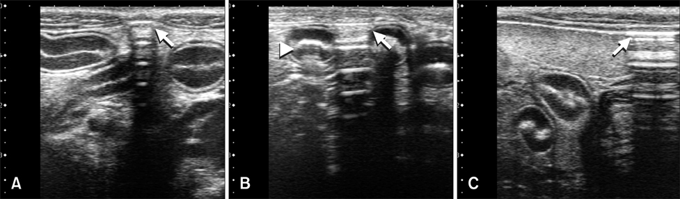
Fig. 1 Ultrasonographic images in the sagittal orientation of the umbilical region (A) and left lumbar (B and C) showing free abdominal air (arrows) that led to increased echogenicity and thickening of the peritoneal stripe with reverberation artifacts. Free abdominal air was present along the ventral peritoneum adjacent to the spleen and small intestine. Intraluminal air was also observed in the intestinal lumen (arrowhead) and associated with "dirty" acoustic shadowing.
Reference
-
1. Asrani A. Sonographic diagnosis of pneumoperitoneum using the 'enhancement of the peritoneal stripe sign.' A prospective study. Emerg Radiol. 2007; 14:29–39.
Article2. Choi H, Lee Y, Park K, Yeon S, Lee H. Sonographic detection of small amounts of free peritoneal gas in Beagle dogs. J Vet Med Sci. 2012; 74:491–494.
Article3. Earls JP, Dachman AH, Colon E, Garrett MG, Molloy M. Prevalence and duration of postoperative pneumoperitoneum: sensitivity of CT vs left lateral decubitus radiography. AJR Am J Roentgenol. 1993; 161:781–785.
Article4. Field S, Chan O. Acute abdomen: abdominal trauma. In : Sutton D, Whitehouse RW, editors. Textbook of Radiology and Imaging. 6th ed. New York: Churchill Livingstone;1998. p. 919–921.5. Grechenig W, Peicha G, Clement HG, Grechenig M. Detection of pneumoperitoneum by ultrasound examination: an experimental and clinical study. Injury. 1999; 30:173–178.
Article6. Hanbidge AE, Lynch D, Wilson SR. US of the peritoneum. Radiographics. 2003; 23:663–685.
Article7. Lee DH, Lim JH, Ko YT, Yoon Y. Sonographic detection of pneumoperitoneum in patients with acute abdomen. AJR Am J Roentgenol. 1990; 154:107–109.
Article8. Muradali D, Wilson S, Burns PN, Shapiro H, Hope-Simpson D. A specific sign of pneumoperitoneum on sonography: enhancement of the peritoneal stripe. AJR Am J Roentgenol. 1999; 173:1257–1262.
Article
- Full Text Links
-
- Actions
-
Cited
- CITED
-
- Close
- Share
-
- Similar articles
-
- Ultrasonographic assessment of experimentally induced gastric perforation in beagle dogs
- Usefulness of transthoracic lung ultrasound for the diagnosis of mild pneumothorax
- ‘Triangular Cord’ Sign in Biliary Atresia
- Endometrial evaluation with transvaginal ultrasonography for the screening of endometrial hyperplasia or cancer in premenopausal and perimenopausal women
- Emphysematous Pyelonephritis Associated with Pneumoperitoneum and Pneumomediastinum: A Case Report
