The effect of fibronectin-coated implant on canine osseointegration
- Affiliations
-
- 1Department of Prosthodontics, Yonsei University College of Dentistry, Seoul, Korea.
- 2Department of Periodontology, Research Institute for Periodontal Regeneration, Yonsei University College of Dentistry, Seoul, Korea. shchoi726@yuhs.ac
- 3Department of Dental Biomaterials Science and Dental Research Institute, Seoul National University School of Dentistry, Seoul, Korea.
- 4Institute of Physics and Applied Physics, Atomic-Scale Surface Research Center, Yonsei University, Seoul, Korea.
- KMID: 1783619
- DOI: http://doi.org/10.5051/jpis.2011.41.5.242
Abstract
- PURPOSE
The purpose of this study was to characterize the osseointegration of the fibronectin-coated implant surface.
METHODS
Sand-blasted, large-grit, acid-etched (SLA) surface implants, with or without a thin calcium phosphate and fibronectin coating, were placed in edentulous mandibles of dogs 8 weeks after extraction. All dogs were sacrificed forhistological and histomorphometric evaluation after 4- and 8-week healing periods.
RESULTS
All types of implants were clinically stable without any mobility. Although the bone-to-implant contact and bone density of the SLA implants coated with calcium phosphate (CaP)/fibronectin were lower than the uncoated SLA implants, there were no significant differences between the uncoated SLA surface group and the SLA surface coated with CaP/fibronectin group.
CONCLUSIONS
Within the limits of this study, SLA surfaces coated with CaP/fibronectin were shown to have comparable bone-to-implant contact and bone density to uncoated SLA surfaces.
Keyword
MeSH Terms
Figure
-

Figure 1 Histologic view of the fibronectin-coated sandblasted, large-grit, acid-etched group at 4 weeks (Goldner's trichrome staining, ×40). Osteoid matrix (arrow) and newly formed mineralized bone in the interthread space were observed.
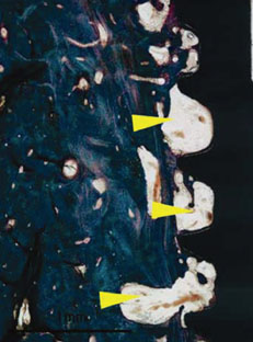
Figure 2 Histologic view of the sandblasted, large-grit, acid-etched group at 4 weeks (Goldner's trichrome staining, ×40). A thin lining of newly formed bone was observed on the implant surface. Marrow space (arrow) near the osteoid matrix was observed.
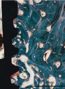
Figure 3 Histologic view of the fibronectin-coated sandblasted, large-grit, acid-etched group at 8 weeks (Goldner's trichrome staining, ×40). Osseointegration of each implant was confirmed. Newly formed mineralized bone in the interthread space was observed.
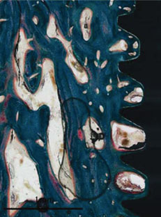
Figure 4 Histologic view of the sandblasted, large-grit, acid-etched group at 8 weeks (Goldner's trichrome staining, ×40). The newly formed bone in the interthread space was in close contact with implant surface without any gaps or dense fibrous connective tissue. No apical epithelial migration was found. No inflammatory cell infiltrate was present around the implants.
Cited by 1 articles
-
Early bone healing onto implant surface treated by fibronectin/oxysterol for cell adhesion/osteogenic differentiation: in vivo experimental study in dogs
Jung-Seok Lee, Jin-Hyuk Yang, Ji-Youn Hong, Ui-Won Jung, Hyeong-Cheol Yang, In-Seop Lee, Seong-Ho Choi
J Periodontal Implant Sci. 2014;44(5):242-250. doi: 10.5051/jpis.2014.44.5.242.
Reference
-
1. Dohan Ehrenfest DM, Coelho PG, Kang BS, Sul YT, Albrektsson T. Classification of osseointegrated implant surfaces: materials, chemistry and topography. Trends Biotechnol. 2010. 28:198–206.
Article2. Atieh MA, Payne AG, Duncan WJ, de Silva RK, Cullinan MP. Immediate placement or immediate restoration/loading of single implants for molar tooth replacement: a systematic review and meta-analysis. Int J Oral Maxillofac Implants. 2010. 25:401–415.3. Bornstein MM, Hart CN, Halbritter SA, Morton D, Buser D. Early loading of nonsubmerged titanium implants with a chemically modified sand-blasted and acid-etched surface: 6-month results of a prospective case series study in the posterior mandible focusing on peri-implant crestal bone changes and implant stability quotient (ISQ) values. Clin Implant Dent Relat Res. 2009. 11:338–347.
Article4. Morton D, Bornstein MM, Wittneben JG, Martin WC, Ruskin JD, Hart CN, et al. Early loading after 21 days of healing of nonsubmerged titanium implants with a chemically modified sandblasted and acid-etched surface: two-year results of a prospective two-center study. Clin Implant Dent Relat Res. 2010. 12:9–17.
Article5. Oates TW, Valderrama P, Bischof M, Nedir R, Jones A, Simpson J, et al. Enhanced implant stability with a chemically modified SLA surface: a randomized pilot study. Int J Oral Maxillofac Implants. 2007. 22:755–760.6. Yoon HJ, Song JE, Um YJ, Chae GJ, Chung SM, Lee IS, et al. Effects of calcium phosphate coating to SLA surface implants by the ion-beam-assisted deposition method on self-contained coronal defect healing in dogs. Biomed Mater. 2009. 4:044107.
Article7. Kokubo T, Kim HM, Kawashita M, Nakamura T. Bioactive metals: preparation and properties. J Mater Sci Mater Med. 2004. 15:99–107.8. de Groot K, Wolke JG, Jansen JA. Calcium phosphate coatings for medical implants. Proc Inst Mech Eng H. 1998. 212:137–147.
Article9. Daculsi G, Laboux O, Malard O, Weiss P. Current state of the art of biphasic calcium phosphate bioceramics. J Mater Sci Mater Med. 2003. 14:195–200.10. Choi JM, Kim HE, Lee IS. Ion-beam-assisted deposition (IBAD) of hydroxyapatite coating layer on Ti-based metal substrate. Biomaterials. 2000. 21:469–473.
Article11. Lee IS, Whang CN, Kim HE, Park JC, Song JH, Kim SR. Various Ca/P ratios of thin calcium phosphate films. Mater Sci Eng C Biomim Mater Sens Syst. 2002. 22:15–20.
Article12. Hynes RO. Fibronectins. Sci Am. 1986. 254:42–51.
Article13. Horbett TA. Chapter 13 Principles underlying the role of adsorbed plasma proteins in blood interactions with foreign materials. Cardiovasc Pathol. 1993. 2:3 Suppl. 137–148.
Article14. Puleo DA, Nanci A. Understanding and controlling the bone-implant interface. Biomaterials. 1999. 20:2311–2321.
Article15. Cannas M, Denicolai F, Webb LX, Gristina AG. Bioimplant surfaces: binding of fibronectin and fibroblast adhesion. J Orthop Res. 1988. 6:58–62.16. Dean JW 3rd, Culbertson KC, D'Angelo AM. Fibronectin and laminin enhance gingival cell attachment to dental implant surfaces in vitro. Int J Oral Maxillofac Implants. 1995. 10:721–728.17. El-Ghannam A, Starr L, Jones J. Laminin-5 coating enhances epithelial cell attachment, spreading, and hemidesmosome assembly on Ti-6A1-4V implant material in vitro. J Biomed Mater Res. 1998. 41:30–40.
Article18. Roehlecke C, Witt M, Kasper M, Schulze E, Wolf C, Hofer A, et al. Synergistic effect of titanium alloy and collagen type I on cell adhesion, proliferation and differentiation of osteoblast-like cells. Cells Tissues Organs. 2001. 168:178–187.
Article19. Sugino A, Miyazaki T, Kawachi G, Kikuta K, Ohtsuki C. Relationship between apatite-forming ability and mechanical properties of bioactive PMMA-based bone cement modified with calcium salts and alkoxysilane. J Mater Sci Mater Med. 2008. 19:1399–1405.
Article20. Chen C, Lee IS, Zhang SM, Yang HC. Biomimetic apatite formation on calcium phosphate-coated titanium in Dulbecco's phosphate-buffered saline solution containing CaCl(2) with and without fibronectin. Acta Biomater. 2010. 6:2274–2281.
Article21. Hayakawa T, Yoshinari M, Nemoto K. Direct attachment of fibronectin to tresyl chloride-activated titanium. J Biomed Mater Res A. 2003. 67:684–688.
Article22. Park JM, Koak JY, Jang JH, Han CH, Kim SK, Heo SJ. Osseointegration of anodized titanium implants coated with fibroblast growth factor-fibronectin (FGF-FN) fusion protein. Int J Oral Maxillofac Implants. 2006. 21:859–866.23. do Serro AP, Fernandes AC, de Jesus Vieira Saramago B. Calcium phosphate deposition on titanium surfaces in the presence of fibronectin. J Biomed Mater Res. 2000. 49:345–352.
Article24. Weiss RE, Reddi AH. Synthesis and localization of fibronectin during collagenous matrix-mesenchymal cell interaction and differentiation of cartilage and bone in vivo. Proc Natl Acad Sci U S A. 1980. 77:2074–2078.
Article25. Saba TM, Jaffe E. Plasma fibronectin (opsonic glycoprotein): its synthesis by vascular endothelial cells and role in cardiopulmonary integrity after trauma as related to reticuloendothelial function. Am J Med. 1980. 68:577–594.
Article26. Tamada Y, Ikada Y. Effect of preadsorbed proteins on cell adhesion to polymer surfaces. J Colloid Interface Sci. 1993. 155:334–339.
Article27. Jönsson U, Ivarsson B, Lundström I, Berghem L. Adsorption behavior of fibronectin on well-characterized silica surfaces. J Colloid Interface Sci. 1982. 90:148–163.
Article28. Malmstrom HS, Fritz ME, Timmis DP, Van Dyke TE. Osseo-integrated implant treatment of a patient with rapidly progressive periodontitis. A case report. J Periodontol. 1990. 61:300–304.
Article29. Mombelli A, Lang NP. The diagnosis and treatment of peri-implantitis. Periodontol 2000. 1998. 17:63–76.
Article30. Davies JE. Mechanisms of endosseous integration. Int J Prosthodont. 1998. 11:391–401.
- Full Text Links
-
- Actions
-
Cited
- CITED
-
- Close
- Share
-
- Similar articles
-
- Teh Effect of Hydroxyapatite Coating on the Mechanical Strengths and Histologic Profiles of Porous Titanium Implants in Dogs
- A comparative experimental study of bone ingrowth and osseointegration in hydroxyapatite-coated vs. porous-coated implants
- The effect of purified human BMP with DLB(hBMP-I) on osseointegration of immediate titanium implants: cases report
- Comparative Study on Osseointegration of Calcium Metaphosphate (CMP) Coated Implant to RBM Implant in the Femur of Rabbits
- Bone apposition on implants coated with calcium phosphate by ion beam assisted deposition in oversized drilled sockets: a histologic and histometric analysis in dogs
