Acquired Omental Cystic Lymphangioma after Subtotal Gastrectomy: A Case Report
- Affiliations
-
- 1Department of Surgery, Korea University College of Medicine, Seoul, Korea. junwonum@korea.ac.kr
- 2Department of Pathology, Korea University College of Medicine, Seoul, Korea.
- KMID: 1783159
- DOI: http://doi.org/10.3346/jkms.2009.24.6.1212
Abstract
- We herein describe a case of cystic lymphangioma in the greater omentum of the remnant stomach, which is thought it to be related with subtotal gastrectomy 10 yr ago for early gastric cancer. A 76-yr-old man was admitted to our department with postprandial abdominal discomfort and bowel habit change. Intraabdominal multilocular cystic mass was detected by ultrasonography and computed tomography. We performed a complete En-bloc tumor resection including spleen and distal pancreas, and histological examination confirmed cystic lymphangioma originated from the greater omentum of the remnant stomach. Although the etiology of omental lymphangioma remains largely unclear, these findings suggested strongly that obstruction of the lymphatic vessels after gastric resection for gastric carcinoma might be the most plausible cause. The surgical extirpation with resection of organs involved appears to be a treatment of choice for such unusual case.
Keyword
MeSH Terms
Figure
-
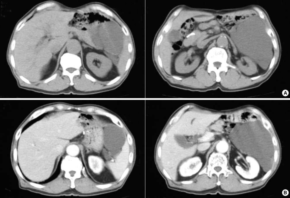
Fig. 1 An abdominal CT scan showed about 12.0×15.0 cm sized non-enhancing cystic mass in the left abdominal cavity. This mass was attached to pancreas body and tail superomedially, and displaced a remnant of stomach medially, spleen laterally, and adjacent bowel loops inferomedially. This cystic mass contained internal septum, like structure in the upper aspect of tumor.

Fig. 2 Gross specimen showed an encapsulated cystic tumor combined with distal pancreas and spleen (A, Anterior view; B, Posterior view).
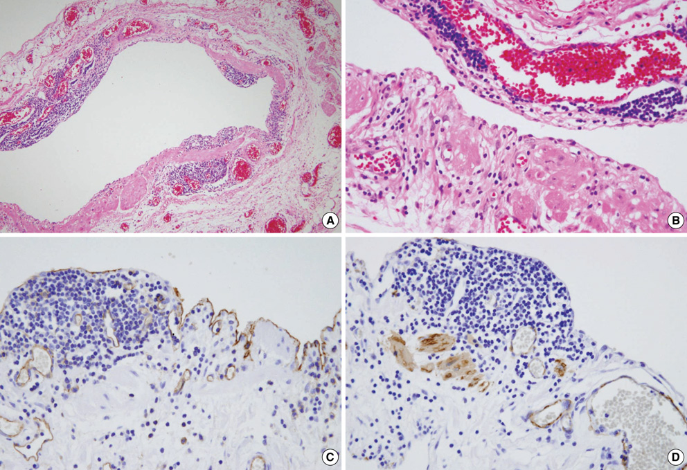
Fig. 3 Histology of lymphangioma. (A, B) Cystic wall was consisted of fibroconnective tissue accompanied by dilated lymphatic spaces and lymphoid cell aggregations in the endothelial lining of lymphatic vessels (H&E ×100, ×400). Immunohistochemical staining. (C) Positive for CD31 in the lining cells of dilated lymphatic vessels (×400). (D) Positive for SMA in the cystic wall, and they were smooth muscle bundles surrounding lymphatic vascular endothelium (×400).
Reference
-
1. Takiff H, Calabria R, Yin L, Stabile BE. Mesenteric cysts and intraabdominal cystic lymphangiomas. Arch Surg. 1985. 120:1266–1269.
Article2. Roisman I, Manny J, Fields S, Shiloni E. Intra-abdominal lymphangioma. Br J Surg. 1989. 76:485–489.
Article3. Fonkalsrud EW. Congenital malformations of the lymphatic system. Semin Pediatr Surg. 1994. 3:62–69.4. de Perrot M, Rostan O, Morel P, Le Coultre C. Abdominal lymphangioma in adults and children. Br J Surg. 1998. 85:395–397.
Article5. Losanoff JE, Richman BW, El-Sherif A, Rider KD, Jones JW. Mesenteric cystic lymphangioma. J Am Coll Surg. 2003. 196:598–603.
Article6. Allen JG, Riall TS, Cameron JL, Askin FB, Hruban RH, Campbell KA. Abdominal lymphangiomas in adults. J Gastrointest Surg. 2006. 10:746–751.
Article7. Chung JH, Suh YL, Park IA, Jang JJ, Chi JG, Kim YI, Kim WH. A pathologic study of abdominal lymphangiomas. J Korean Med Sci. 1999. 14:257–262.
Article8. Miettinen M, Lindenmayer AE, Chaubal A. Endothelial cell markers cd31, cd34, and bnh9 antibody to h- and y-antigens--evaluation of their specificity and sensitivity in the diagnosis of vascular tumors and comparison with von willebrand factor. Mod Pathol. 1994. 7:82–90.
- Full Text Links
-
- Actions
-
Cited
- CITED
-
- Close
- Share
-
- Similar articles
-
- A Acquired Lymphangioma Circumscriptum of the Nipple Areola
- Cystic Lymphangioma of Rectum-A Case Report and Review of Literature
- Retroperitoneal cystic lymphangioma in an aged man: report of a case and review of the literature
- Cystic lymphangioma of the pancreas: a case report
- Cystic Lymphangioma Involving the Mesentery and the Retroperitoneum: A Case Report
