High Resolution Time Resolved Contrast Enhanced MR Angiography Using k-t FOCUSS
- Affiliations
-
- 1Bio-Imaging & Signal Processing Lab, Department of Bio/Brain Engineering, Korea Advanced Institute of Science & Technology (KAIST), Korea. jong.ye@kaist.ac.kr
- 2Department of Radiology and Research Institute of Radiological Science, Yonsei University College of Medicine, Korea.
- KMID: 1782989
- DOI: http://doi.org/10.13104/jksmrm.2010.14.1.10
Abstract
- PURPOSE
Recently, the Recon Challenge at the 2009 ISMRM workshop on Data Sampling and Image Reconstruction at Sedona, Arizona was held to evaluate feasibility of highly accelerated acquisition of time resolved contrast enhanced MR angiography. This paper provides the step-by-step description of the winning results of k-t FOCUSS in this competition.
MATERIALS AND METHODS
In previous works, we proved that k-t FOCUSS algorithm successfully solves the compressed sensing problem even for less sparse cardiac cine applications. Therefore, using k-t FOCUSS, very accurate time resolved contrast enhanced MR angiography can be reconstructed. Accelerated radial trajectory data were synthetized from X-ray cerebral angiography images and provided by the organizing committee, and radiologists double blindly evaluated each reconstruction result with respect to the ground-truth data.
RESULTS
The reconstructed results at various acceleration factors demonstrate that each components of compressed sensing, such as sparsifying transform and incoherent sampling patterns, etc can have profound effects on the final reconstruction results.
CONCLUSION
From reconstructed results, we see that the compressed sensing dynamic MR imaging algorithm, k-t FOCUSS enables high resolution time resolved contrast enhanced MR angiography.
Keyword
MeSH Terms
Figure
-

Fig. 1 Time resolved CE-MRA can be spasified using FT or KLT. Unlike FT, KLT is data dependent transform so that KLT bases should be obtained from given data set. The proposed method first applied k-t FOCUSS with temporal FT for initial reconstruction. Then, by thresholding the temporal averaged image, a mask is obtained and KLT bases are estimated from the temporal variation at the mask location. After KLT, the coefficients are usually very sparse.
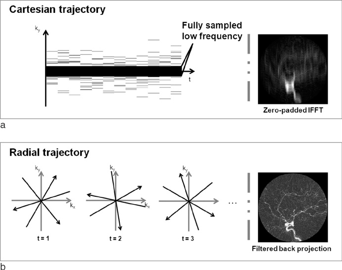
Fig. 2 Randomly sampled Cartesian and radial trajectories are illustrated with reconstructed images using conventional methods in (a) and (b), respectively.

Fig. 3 Two different time frames representing spread of bolus are illustrated to compare the performance of FT without up-sampling, FT with up-sampling, and KLT with up-sampling. The acceleration factor was 12 and up-sampling factor was 2. Without up-sampling, streaking and aliasing artifacts are visible as indicated by arrows. Up-sampling improves the image quality. Using KLT, further improved images can be reconstructed. KLT results show the finest vessel structure in magnified images.
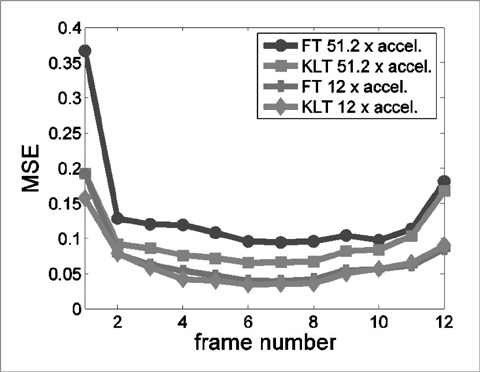
Fig. 4 Mean square error (MSE) was plotted for FT and KLT at 51.2 and 12-fold acceleration. KLT shows significantly reduced MSE especially at high acceleration compared to FT.
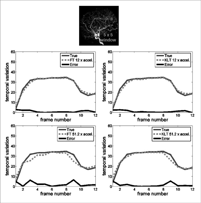
Fig. 5 Temporal variation of averaged pixel values within the specified window is plotted for FT and KLT at 12 and 51.2-fold accelerations. At 12-fold acceleration, both methods show accurate time variations. However, at 51.2-fold acceleration, only KLT method correctly follows true temporal variation.

Fig. 6 k-t FOCUSS with KLT was implemented in Cartesian and radial trajectories at 12-fold acceleration and both resultant images are compared in (a). Both results look similar but in magnified version radial trajectory shows finer vessel structures. Similarly, in y-axis and t-axis plot in (b) and (c), radial trajectory shows more accurate results.
Reference
-
1. Urban B, Ratner L, Fishman E. Three-dimensional Volume-rendered CT Angiography of the Renal Arteries and Veins: Normal Anatomy, Variants, and Clinical Application 1. Radiographics. 2001. 21(2):373–386.2. Zanzonico P, Rothenberg L, Strauss H. Radiation Exposure of Computed Tomography and Direct Intracoronary Angiography Risk has its Reward. J Am Coll Cardiol. 2006. 47(9):1846–1849.3. Lee VS. Cardiovascular MRI: Physical principles to practical protocols. 2006. Philadelpia: Lippincott Williams & Wilkins.4. Pruessmann KP, Weigher M, Scheidegger MB, Boesiger P. SENSE: Sensitivity encoding for fast MRI. Magn Reson Med. 1999. 42(5):952–962.5. Sodickson DK, Manning WJ. Simultaneous acquisition of spatial harmonics (SMASH): fast imaging with radiofrequency coil arrays. Magn Reson Med. 1997. 38(4):591–603.6. Griswold MA, Jakob PM, Heidemann RM, Nittka M, Jellus V, Wang J, Kiefer B, Haase A. Generalized autocalibrating partially parallel acquisitions (GRAPPA). Magn Reson Med. 2002. 47(6):1202–1210.7. Sodickson D, McKenzie C, Li W, Wolff S, Manning W, Edelman R. "Contrast-enhanced 3D MR Angiography with Simultaneous Acquisition of Spatial Harmonics: A Pilot Study 1. Radiology. 2000. 217(1):284–289.8. Wilson GJ, Hoogeveen R, Willinek W, Muthupillai R, Maki J. "Parallel Imaging in MR Angiography". Top Magn Reson Imaging. 2004. 15(3):169–185.9. Lustig M, Donoho D, Pauly J. Sparse MRI: The application of compressed sensing for rapid MR imaging. Magn Reson Med. 2007. 58(6):1182–1195.10. Donoho DL. Compressed sensing. IEEE Trans Inf Theory. 2006. 52(5):1289–1306.11. Jung H, Ye JC, Kim EY. Improved k-t BLAST and k-t SENSE using FOCUSS. Phys Med Biol. 2007. 52(11):3201–3226.12. Jung H, Sung K, Nayak KS, Kim EY, Ye JC. k-t FOCUSS: a general compressed sensing framework for high resolution dynamic MRI. Magn Reson Med. 2009. 61:103–116.13. Winkelmann S, Schaeffter T, Koehler T, Eggers H, Doessel O. An optimal radial profile order based on the Golden Ratio for time-resolved MRI. IEEE Trans Med Imaging. 2007. 26(1):68–76.14. Poor HV. An Introduction of Signal Detection and Estimation. 1994. 2nd ed. New York: Springer-Verlag.15. Liang Z. Spatiotemporal Imaging with Partially Separable Functions. 2007. In : 4th IEEE International Symposium on Biomedical Imaging: From Nano to Macro; 988–991.16. Mistretta C, Wieben O, Velikina J, Block W, Perry J, Wu Y, Johnson K, Wu Y. Highly constrained backprojection for time-resolved MRI. Magn Reson Med. 2006. 55(1):30–40.17. O'Halloran RL, Wen Z, Holmes JH, Fain SB. Iterative projection reconstruction of time-resolved images using highly-constrained back-proejction (HYPR). Magn Reson Med. 2008. 59(1):132–139.18. Wild J, Paley M, Kasuboski L, Swift A, Fichele S, Woodhouse N, Griffiths P, van Beek E. Dynamic radial projection MRI of inhaled hyperpolarized 3 He gas. Magn Reson Med. 2003. 49(6):991–997.19. Liang ZP, Lauterbur PC. Principles of magnetic resonance imaging: A signal processing perspective. 2000. New York: IEEE press.
- Full Text Links
-
- Actions
-
Cited
- CITED
-
- Close
- Share
-
- Similar articles
-
- Contrast Enhanced MR Angiography after Metallic Stent Placement: Experimental Study
- High field strength magnetic resonance imaging of abdominal diseases
- Subtraction MR Venography Acquired from Time-Resolved Contrast-Enhanced MR Angiography: Comparison with Phase-Contrast MR Venography and Single-Phase Contrast-Enhanced MR Venography
- Ultrafast Contrast-Enhanced MR Angiography of the Carotid Artery: Time Optimization for Discrimination of theArterial from the Venous Phase
- Dynamic Contrast-Enhanced T2*-Weighted Imaging in Acute Cerebral Infarction: Usefulness in Assessment of Cerebral Hemodynamics
