Yonsei Med J.
2010 Nov;51(6):984-986. 10.3349/ymj.2010.51.6.984.
Variation of the Infrahyoid Muscle: Duplicated Omohyoid and Appearance of the Levator Glandulae Thyroideae Muscles
- Affiliations
-
- 1Department of Anatomy, Catholic Institution for Applied Anatomy, College of Medicine, The Catholic University of Korea, Seoul, Korea.
- 2Department of Anatomy, Kwandong University College of Medicine, Gangneung, Korea. kslee@kd.ac.kr
- KMID: 1779653
- DOI: http://doi.org/10.3349/ymj.2010.51.6.984
Abstract
- The embryologic origin of the omohyoid muscle is different from that of the other neck muscles. A number of variations such as the absence of muscle, variable sites of origin and insertion, and multiple bellies have been reported. However, variations in the inferior belly of the omohyoid muscle are rare. There have been no reports of the combined occurrence of the omohyoid muscle variation with the appearance of the levator glandulase thyroideae muscle. Routine dissection of a 51-year-old female cadaver revealed a duplicated omohyoid muscle and the appearance of the levator glandulae thyroideae muscle. In this case, the two inferior bellies of the omohyoid muscle were found to originate inferiorly from the superior border of the scapula. One of the inferior bellies generally continued to the superior belly with the tendinous intersection. The other inferior belly continued into the sternohyoid muscle without the tendinous intersection. In this case, the levator glandulae thyroideae muscle appeared on the left side, which attached from the upper border of the thyroid gland to the inferior border of the thyroid cartilage. These variations are significant for clinicians during endoscopic diagnosis and surgery because of the arterial and nervous damage due to iatrogenic injuries. The embryologic origins of the omohyoid and levator glandulae thyroideae muscles may be similar based on the descriptions in the relevant literature.
MeSH Terms
Figure
-
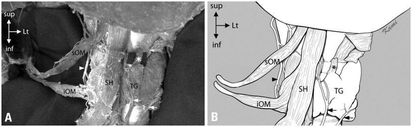
Fig. 1 Variations of the infrahyoid muscles. (A) Photograph of variations of the infrahyoid muscles. (B) Sschematic drawing of these variations. sOM, superior omohyoid muscle; iOM, inferior omohyoid muscle; SH, sternohyoid muscle; TG, thyroid gland; asterisk. *levator glandulae thyroideae muscle; arrow head, ansa cervicalis. arrow, inferior thyroid vein.
Reference
-
1. Hatipoğlu ES, Kervancioğlu P, Tuncer MC. An unusual variation of the omohyoid muscle and review of literature. Ann Anat. 2006. 188:469–472.
Article2. Rai R, Nayak SR, Ranade AV, Prabhu LV, Vadgaonkar R. Duplicated omohyoid muscle and its clinical significance. Rom J Morphol Embryol. 2007. 48:295–297.3. Gray H, Williams PL, Bannister LH. Gray's anatomy: The anatomical basis of clinical practice. 2006. 39th ed. New York: Churchill Livingstone.4. Winslow JB. Exposition anatomique de la structure du corps humain. 1743. Amsterdam: Wetstein.5. Sukekawa R, Itoh I. Anatomical study of the human omohyoid muscle: regarding intermediate morphologies between normal and anomalous morphologies of the superior belly. Anat Sci Int. 2006. 81:107–114.6. Loth E. Anthropologie des parties Molles. 1931. Paris: Werner Wachsmuth.7. Lehr RP Jr. Musculus levator glandulae thyroideae: an observation. Anat Anz. 1979. 146:494–496.8. Loukas M, Merbs W, Tubbs RS, Curry B, Jordan R. Levator glandulae thyroideae muscle with three slips. Anat Sci Int. 2008. 83:273–276.9. Mori M. Statistics on the musculature of the Japanese. Okajimas Folia Anat Jpn. 1964. 40:195–300.
Article10. Tubbs RS, Salter EG, Oakes WJ. Unusual origin of the omohyoid muscle. Clin Anat. 2004. 17:578–582.
Article11. Shih TY, Chuang JH. Fibrosis of the omohyoid muscle--an unusual cause of torticollis. J Pediatr Surg. 1998. 33:741–742.
Article12. Patra P, Gunness TK, Robert R, Rogez JM, Heloury Y, Le Hur PA, et al. Physiologic variations of the internal jugular vein surface, role of the omohyoid muscle, a preliminary echographic study. Surg Radiol Anat. 1988. 10:107–112.
Article13. Krishnan KG, Pinzer T, Reber F, Schackert G. Endoscopic exploration of the brachial plexus: technique and topographic anatomy--a study in fresh human cadavers. Neurosurgery. 2004. 54:401–408.
Article14. Kojima H, Hirano S, Shoji K, Omori K, Honjo I. Omohyoid muscle transposition for the treatment of bowed vocal fold. Ann Otol Rhinol Laryngol. 1996. 105:536–540.
Article15. Crumley RL. Muscle transfer for laryngeal paralysis. Restoration of inspiratory vocal cord abduction by phrenic-omohyoid transfer. Arch Otolaryngol Head Neck Surg. 1991. 117:1113–1117.
Article16. Watanabe S, Suda M. On the musculus levator glandulae thyroideae of the Ainu. Saporro Igacu Zasshi. 1962. 21:115–120.17. Harjeet A, Sahni D, Jit I, Aggarwal AK. Shape, measurements and weight of the thyroid gland in northwest Indians. Surg Radiol Anat. 2004. 26:91–95.
Article18. Gregory JK, Guse DM. Unique variant of levator glandulae thyroideae muscle. Clin Anat. 2007. 20:966–967.
Article19. Eisler P, Der M. Levator glandulae thyroideae und verwandte praelaryngeale Muskelbildungen. Anat Anz. 1900. 17:183–189.
- Full Text Links
-
- Actions
-
Cited
- CITED
-
- Close
- Share
-
- Similar articles
-
- Appearance of the Cleidohyoideus Muscle Combined with the Multiple Variations of the Infrahyoid Muscle
- Unusual morphology of the superior belly of omohyoid muscle
- Unusual muscle of the anterior neck: cadaveric findings with surgical applications
- Omohyoid Muscle Syndrome
- Pharyngeal Closure Reinforced With Sternohyoid and Omohyoid Muscle After Total Laryngectomy
