Granular Cell Tumor of the Descending Colon Treated by Endoscopic Mucosal Resection: A Case Report and Review of the Literature
- Affiliations
-
- 1Department of Internal Medicine, Kyunghee University College of Medicine, Seoul, Korea. dramc@hanmail.net
- 2Department of Pathology, Kyunghee University College of Medicine, Seoul, Korea.
- KMID: 1779142
- DOI: http://doi.org/10.3346/jkms.2009.24.2.337
Abstract
- Although colorectal granular cell tumors (GCTs) are rare, their incidental finding has increased as the use of diagnostic colonoscopy has become more common. Here we describe the case of a 41-yr-old man with a GCT in the descending colon that was detected after a screening colonoscopy. Endoscopic examination revealed a yellowish submucosal tumor, 13x12 mm in diameter, in the descending colon. Endoscopic mucosal resection (EMR) followed by histological examination revealed that the tumor was composed of plump histiocyte-like cells with an abundant granular eosinophilic cytoplasm and small round nuclei. The tumor cells expressed S-100 protein and stained with periodic acid-Schiff, but were negative for desmin and cytokeratin. The resected tumor was diagnosed as a GCT. Colonoscopists should consider the possibility of GCT in the differential diagnosis of yellowish submucosal tumors of the colon. In such patients, EMR seems to be a feasible and safe approach for diagnosis and treatment.
MeSH Terms
Figure
-

Fig. 1 Colonoscopy detected an approximately 13×12 mm yellowish, submucosal tumor in the descending colon. It was hard in consistency without ulceration.

Fig. 2 Histological findings of the tumor. (A) The resected tumor was covered with normal mucosa (H&E, ×20). A nested growth of nonuniform large tumor cells with slightly pleomorphic nuclei (inlet, H&E, ×400). (B) Some granules were positive for periodic acid-Schiff (PAS, ×400).
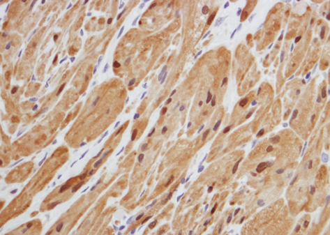
Fig. 3 Histological findings of the tumor showing positive immunoreaction for S-100 protein (immunohistochemical stain, ×400).
Cited by 1 articles
-
A Case of Malignant Granular Cell Tumor in the Sigmoid Colon
Sang Myung Choi, Seung Goun Hong, Shin Myung Kang, Byung Gi Chae, Sung Jin Kim, Pyung Kang Park, Hyun Sung Park
Clin Endosc. 2014;47(2):197-200. doi: 10.5946/ce.2014.47.2.197.
Reference
-
1. Lack EE, Worsham GF, Callihan MD, Crawford BE, Klappenbach S, Rowden G, Chun B. Granular cell tumor: a clinicopathologic study of 110 patients. J Surg Oncol. 1980. 13:301–316.
Article2. Kulaylat MN, King B. Granular cell tumor of the colon. Dis Colon Rectum. 1996. 39:711.
Article3. Yasuda I, Tomita E, Nagura K, Nishigaki Y, Yamada O, Kachi H. Endoscopic removal of granular cell tumors. Gastrointest Endosc. 1995. 41:163–167.
Article4. Melo CR, Melo IS, Schmitt FC, Fagundes R, Amendola D. Multicentric granular cell tumor of the colon: report of a patient with 52 tumors. Am J Gastroenterol. 1993. 88:1785–1787.5. Joshi A, Chandrasoma P, Kiyabu M. Multiple granular cell tumor of the gastrointestinal tract with subsequent development of esophageal squamous cell carcinoma. Dig Dis Sci. 1992. 37:1612–1618.6. Johnston J, Helwig EB. Granular cell tumors of the gastrointestinal tract and perianal region: a study of 74 cases. Dig Dis Sci. 1981. 26:807–816.
Article7. Armin A, Connelly EM, Rowden G. An immunoperoxidase investigation of S-100 protein in granular cell myoblastomas: evidence for Schwann cell derivation. Am J Clin Pathol. 1983. 79:37–44.
Article8. Choi JK, Choi MG, Choi KY, Chung IS, Cha SB, Chung KW, Sun HS, Kim BS, Choi YJ, Lee AH. A case of colonoscopically removed granular cell tumor in the ascending colon. Korean J Gastrointest Endosc. 1991. 11:383–386.9. Yamada T, Fujiwara Y, Sasatomi E, Nakano S, Tokunaga O. Granular cell tumor in the ascending colon. Intern Med. 1995. 34:657–660.
Article10. Nakachi A, Miyazato H, Oshiro T, Shimoji H, Shiraishi M, Muto Y. Granular cell tumor of the rectum: a case report and review of the literature. J Gastroenterol. 2000. 35:631–634.
Article11. Rossi GB, de Bellis M, Marone P, Chiara AD, Losito S, Tempesta A. Granular cell tumor of the colon: report of a case and review of the literature. J Clin Gastroenterol. 2000. 30:197–199.12. Kim HS, Cho KA, Hwang DY, Kim KU, Kang YW, Park WK, Yoon SG, Lee KR, Lee JK, Lee JD, Kim KY. A case of granular cell tumor in the appendix. Korean J Gastroenterol. 2000. 36:404–407.13. Irisawa A, Hernandez LV, Bhutani MS. Endosonographic features of a granular cell tumor of the colon. J Ultrasound Med. 2001. 20:1241–1243.
Article14. Endo S, Hirasaki S, Doi T, Endo H, Nishina T, Moriwaki T, Nakauchi M, Masumoto T, Tanimizu M, Hyodo I. Granular cell tumor occurring in the sigmoid colon treated by endoscopic mucosal resection using a transparent cap (EMR-C). J Gastroenterol. 2003. 38:385–389.
Article15. Kim DH, Kim YH, Kwon NH, Song BG, Jung JH, Kim MH, Rhee PL, Kim JJ, Rhee JC. A case of granular cell tumor in the rectum. Korean J Gastrointest Endosc. 2003. 27:88–91.16. Lee SH, Kim SH, Kim BR, Kim HJ, Bhandari S, Jung IS, Hong SJ, Ryu CB, Kim JO, Cho JY, Lee JS, Lee MS, Shim CS, Kim BS, Jin SY. Granular cell tumor of the ascending colon: report of a case. Intestinal Research. 2003. 1:59–63.17. Ryu JH, Choi MH, Kim GS, Choi CS, Suh YA, Jang HJ, Eun CS, Kae SH, Lee J. A case of granular cell tumor of the ascending colon. Korean J Gastrointest Endosc. 2003. 26:439–442.18. Sohn DK, Choi HS, Chang YS, Huh JM, Kim DH, Kim DY, Kim YH, Chang HJ, Jung KH, Jeong SY. Granular cell tumor of the colon: report of a case and review of literature. World J Gastroenterol. 2004. 10:2452–2454.19. Mori T, Orikasa H, Shigematsu T, Yamazaki K. An ultrastructural and immunohistochemical study of a combined submucosal granular cell tumor and lipoma of the colon showing a unique nodule-in-nodule structure: putative implication of CD34 or prominin-2-positive stromal cells in its histopathogenesis. Virchows Arch. 2006. 449:137–139.
Article20. Park NY, Kim KJ, Kim YJ, Roh JH, Im DG, Nam JH, Monn W, Park IP, Park SJ, Cheon BK. A case of granular cell tumor of the colon treated by colonoscopy. Korean J Gastrointest Endosc. 2006. 32:67–70.21. Parfitt JR, McLean CA, Joseph MG, Streutker CJ, Al-Haddad S, Driman DK. Granular cell tumours of the gastrointestinal tract: expression of nestin and clinicopathological evaluation of 11 patients. Histopathology. 2006. 48:424–430.
Article22. Shimoyama M, Sakai Y, Takaku H, Takii Y, Okamoto H, Suda T, Hatakeyama K. A case of multiple granular cell tumors in the cecum. Gastroenterol Endosc. 1999. 41:1330–1335.23. Palazzo L, Landi B, Cellier C, Roseau G, Chaussade S, Couturier D, Barbier J. Endosonographic features of esophageal granular cell tumors. Endoscopy. 1997. 29:850–853.24. Mittal KR, True LD. Origin of granules in granular cell tumor: intracellular myelin formation with autodigestion. Arch Pathol Lab Med. 1988. 112:302–303.25. Uzoaru I, Firfer B, Ray V, Hubbard-Shepard M, Rhee H. Malignant granular cell tumor. Arch Pathol Lab Med. 1992. 116:206–208.26. Matsumoto H, Kojima Y, Inoue T, Takegawa S, Tsuda H, Kobayashi A, Watanabe K. A malignant granular cell tumor of the stomach: report of a case. Surg Today. 1996. 26:119–122.
Article27. Fanburg-Smith JC, Meis-Kindblom JM, Fante R, Kindblom LG. Malignant granular cell tumor of soft tissue: diagnostic criteria and clinicopathologic correlation. Am J Surg Pathol. 1998. 22:779–794.
- Full Text Links
-
- Actions
-
Cited
- CITED
-
- Close
- Share
-
- Similar articles
-
- Granular Cell Tumors of the Cecum: Report of Two Cases and Review of Literature
- Two Granular Cell Tumors of the Colon with the Endosonographic Features
- A Case Of Granular Cell Tumor In The Stomach
- A Case of Granular Cell Tumor of the Ascending Colon
- A Case of Colonoscopically Removed Granular Cell Tumor in the Ascending Colon
