J Korean Med Sci.
2011 Aug;26(8):1052-1060. 10.3346/jkms.2011.26.8.1052.
Relationship between Coronary Artery Calcium Score by Multidetector Computed Tomography and Plaque Components by Virtual Histology Intravascular Ultrasound
- Affiliations
-
- 1The Heart Center, Chonnam National University Hospital, Gwangju, Korea. hyj200@hanmail.net
- 2College of Nursing, Chonnam National University, Gwangju, Korea.
- KMID: 1777844
- DOI: http://doi.org/10.3346/jkms.2011.26.8.1052
Abstract
- The aim of this study was to evaluate the relationship between coronary artery calcium score (CACS) assessed by multidetector computed tomography (MDCT) and plaque components assessed by virtual histology-intravascular ultrasound (VH-IVUS) in 172 coronary artery disease (CAD) patients with 250 coronary lesions. CACS was assessed according to Agatston scoring method by MDCT and patients were divided into four groups: Group I (CACS = 0 [n = 52]); Group II (CACS = 1-100 [n = 99]); Group III (CACS = 101-400 [n = 84]); and Group IV (CACS > 400 [n = 15]). Total atheroma volume was greatest in Group IV (152 +/- 132 microL vs 171 +/- 114 microL vs 195 +/- 149 microL vs 321+/-182 microL, P < 0.001). The absolute dense calcium (DC) and necrotic core (NC) volumes were greatest, and relative DC volume was greatest in Group IV (5.5 +/- 6.6 microL vs 11.0 +/- 10.3 microL vs 15.6 +/- 13.6 microL vs 36.6 +/- 18.2 microL, P < 0.001, and 14.8 +/- 18.2 microL vs 19.5 +/- 18.9 microL vs 22.5 +/- 19.1 microL vs 41.7 +/- 27.9 microL, P < 0.001, and 6.4 +/- 5.3% vs 11.0 +/- 6.2% vs 14.0 +/- 6.5% vs 20.0 +/- 7.8%, P < 0.001, respectively). The absolute plaque and DC and NC volumes and the relative DC volume correlated positively with calcium score. CAD patients with high calcium score have more vulnerable plaque components (greater DC and NC-containing plaques) than those with low calcium score.
Keyword
MeSH Terms
-
Adult
Aged
Calcinosis/*diagnosis/radiography/ultrasonography
Calcium/*analysis
Coronary Angiography
Coronary Artery Disease/*diagnosis/radiography/ultrasonography
Coronary Vessels/pathology
Female
Humans
Male
Middle Aged
*Multidetector Computed Tomography
Necrosis
Plaque, Atherosclerotic/*pathology
*Ultrasonography, Interventional
Figure
-
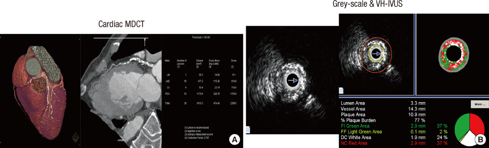
Fig. 1 The examples of coronary artery calcium detected by cardiac multidetector computed tomography (MDCT) (A) and plaque components assessed by virtual histology-intravascular ultrasound (VH-IVUS) (B).
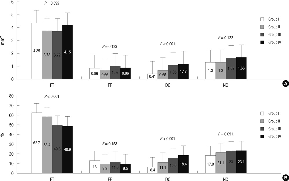
Fig. 2 Absolute (A) and relative (B) plaque components at the minimum lumen area sites. Absolute dense calcium and relative dense calcium (%) areas were greatest in Group IV; in contrast, relative fibrous (%) area was smallest in Group IV.

Fig. 3 Absolute (A) and relative (B) plaque components by volumetric analysis. Absolute and relative dense calcium (%) and necrotic core (%) volumes were greatest in Group IV; in contrast, relative fibrous (%) volume was smallest in Group IV.
Reference
-
1. Javadrashid R, Salehi A, Tarzamni MK, Aslanabadi N, Pak N. Diagnostic efficacy of coronary calcium score in the assessment of significant coronary artery stenosis. Kardiol Pol. 2010. 68:285–291.2. Kitamura A, Kobayashi T, Ueda K, Okada T, Awata N, Sato S, Shimamoto T. Evaluation of coronary artery calcification by multi-detector row computed tomography for the detection of coronary artery stenosis in Japanese patients. J Epidemiol. 2005. 15:187–193.3. Shaw LJ, Raggi P, Schisterman E, Berman DS, Callister TQ. Prognostic value of cardiac risk factors and coronary artery calcium screening for all-cause mortality. Radiology. 2003. 228:826–833.4. Thompson GR, Partridge J. Coronary calcification score: the coronary-risk impact factor. Lancet. 2004. 363:557–559.5. Mitsutake R, Niimura H, Miura S, Zhang B, Iwata A, Nishikawa H, Kawamura A, Kumagai K, Shirai K, Matsunaga A, Saku K. Clinical significance of the coronary calcification score by multidetector row computed tomography for the evaluation of coronary stenosis in Japanese patients. Circ J. 2006. 70:1122–1127.6. LaMonte MJ, FitzGerald SJ, Church TS, Barlow CE, Radford NB, Levine BD, Pippin JJ, Gibbons LW, Blair SN, Nichamar MZ. Coronary artery calcium score and coronary heart disease events in an large cohort of asymptomatic men and women. Am J Epidemiol. 2005. 162:421–429.7. Detrano R, Guerci AD, Carr JJ, Bild DE, Burke G, Folsom AR, Liu K, Shea S, Szklo M, Bluemke DA, O'Leary DH, Tracy R, Watson K, Wong ND, Kronmal RA. Coronary calcium as a predictor of coronary events in four racial or ethnic groups. N Engl J Med. 2008. 358:1336–1345.8. Wong ND, Hsu JC, Detrano RC, Diamond G, Eisenberg H, Gardin JM. Coronary artery calcium evaluation by electron beam computed tomography and its relation to new cardiovascular events. Am J Cardiol. 2000. 86:495–498.9. Baumgart D, Schmermund A, Goerge G, Haude M, Ge J, Adamzik M, Sehnert C, Altmaier K, Groenemeyer D, Seibel R, Erbel R. Comparison of electron beam computed tomography with intracoronary ultrasound and coronary angiography for detection of coronary atherosclerosis. J Am Coll Cardiol. 1997. 30:57–64.10. Funabashi N, Misumi K, Ohnishi H, Asano M, Komuro I. Characterization and morphology of atherosclerotic plaque of coronary arteries: utility of electron-beam tomography to detect non-calcified plaque: a comparison with conventional coronary angiography and intravascular ultrasound. Int J Cardiol. 2007. 115:108–113.11. Henneman MM, Schuijf JD, Pundziute G, van Werkhoven JM, van der Wall EE, Jukema JW, Bax JJ. Non-invasive evaluation with multislice computed tomography in suspected acute coronary syndrome: plaque morphology on multislice computed tomography versus coronary calcium score. J Am Coll Cardiol. 2008. 52:216–222.12. Roberts WL, Moulton L, Law TC, Farrow G, Cooper-Anderson M, Savory J, Rifai N. Evaluation of nine automated high-sensitivity C-reactive protein methods: implications for clinical and epidemiological applications. Part 2. Clin Chem. 2001. 47:418–425.13. Reiber JH, van der Zwet PM, Koning G, von Land CD, van Meurs B, Gerbrands JJ, Buis B, van Voorthuisen AE. Accuracy and precision of quantitative digital coronary arteriography: observer-, short-, and medium-term variabilities. Cathet Cardiovasc Diagn. 1993. 28:187–198.14. Agatston AS, Janowitz WR, Hildner FJ, Zusmer NR, Viamonte M Jr, Detrano R. Quantification of coronary artery calcium using ultrafast computed tomography. J Am Coll Cardiol. 1990. 15:827–832.15. Mintz GS, Nissen SE, Anderson WD, Bailey SR, Erbel R, Fitzgerald PJ, Pinto FJ, Rosenfield K, Siegel RJ, Tuzcu EM, Yock PG. American College of Cardiology Clinical Expert Consensus Document on Standards for Acquisition, Measurement and Reporting of Intravascular Ultrasound Studies (IVUS). A report of the American College of Cardiology Task Force on Clinical Expert Consensus Documents. J Am Coll Cardiol. 2001. 37:1478–1492.16. Nair A, Kuban BD, Tuzcu EM, Schoenhagen P, Nissen SE, Vince DG. Coronary plaque classification with intravascular ultrasound radiofrequency data analysis. Circulation. 2002. 106:2200–2206.17. Ho JS, Fitzgerald SJ, Stolfus LL, Wade WA, Reinhardt DB, Barlow CE, Cannaday JJ. Relation of a coronary artery calcium score higher than 400 to coronary stenoses detected using multidetector computed tomography and to traditional cardiovascular risk factors. Am J Cardiol. 2008. 101:1444–1447.18. Church TS, Levine BD, McGuire DK, Lamonte MJ, Fitzgerald SJ, Cheng YJ, Kimball TE, Blair SN, Gibbons LW, Nichaman MZ. Coronary artery calcium score, risk factors, and incident coronary heart disease events. Atherosclerosis. 2007. 190:224–231.19. Nicholls SJ, Tuzcu EM, Wolski K, Sipahi I, Schoenhagen P, Crowe T, Kapadia SR, Hazen SL, Nissen SE. Coronary artery calcification and changes in atheroma burden in response to established medical therapies. J Am Coll Cardiol. 2007. 49:263–270.20. Okabe T, Mintz GS, Weigold WG, Roswell R, Joshi S, Lee SY, Lee B, Steinberg DH, Roy P, Slottow TL, Kaneshige K, Torguson R, Xue Z, Satler LF, Kent KM, Pichard AD, Weissman NJ, Lindsay J, Waksman R. The predictive value of computed tomography calcium scores: a comparison with quantitative volumetric intravascular ultrasound. Cardiovasc Revasc Med. 2009. 10:30–35.21. Burke AP, Kolodgie FD, Zieske A, Fowler DR, Weber DK, Varghese PJ, Farb A, Virmani R. Morphologic findings of coronary atherosclerotic plaques in diabetics: a postmortem study. Arterioscler Thromb Vasc Biol. 2004. 24:1266–1271.22. Schmermund A, Erbel R. Unstable coronary plaque and its relation to coronary calcium. Circulation. 2001. 104:1682–1687.23. Pitt B, Rubenfire M. Risk stratification for the detection of preclinical coronary artery disease. Circulation. 1999. 99:2610–2612.24. Raggi P, Callister TQ, Cooil B, He ZX, Lippolis NJ, Russo DJ, Zelinger A, Mahmarian JJ. Identification of patients at increased risk of first unheralded acute myocardial infarction by electron-beam computed tomography. Circulation. 2000. 101:850–855.25. Arad Y, Spadaro LA, Goodman K, Newstein D, Guerci AD. Prediction of coronary events with electron beam computed tomography. J Am Coll Cardiol. 2000. 36:1253–1260.26. Fujii K, Carlier SG, Mintz GS, Takebayashi H, Yasuda T, Costa RA, Moussa I, Dangas G, Mehran R, Lansky AJ, Kreps EM, Collins M, Stone GW, Moses JW, Leon MB. Intravascular ultrasound study of patterns of calcium in ruptured coronary plaques. Am J Cardiol. 2005. 96:352–357.27. Ehara S, Kobayashi Y, Kataoka T, Yoshiyama M, Ueda M, Yoshikawa J. Quantification of coronary calcification by intravascular ultrasound. Circ J. 2007. 71:530–535.28. Harada K, Amano T, Uetani T, Funahashi H, Arai K, Okada K, Hirashiki A, Hayashi M, Oshima S, Ishii H, Izawa H, Matsubara T, Murohara T. Accuracy of 64-slice multidetector computed tomography for classification and quantitation of coronary plaque: comparison with integrated backscatter intravascular ultrasound. Int J Cardiol. 2011. 149:95–101.29. Carrascosa PM, Capuñay CM, Garcia-Merletti P, Carrascosa J, Garcia MF. Characterization of coronary atherosclerotic plaques by multidetector computed tomography. Am J Cardiol. 2006. 97:598–602.30. Galonska M, Ducke F, Kertesz-Zborilova T, Meyer R, Guski H, Knollmann FD. Characterization of atherosclerotic plaques in human coronary arteries with 16-slice multidetector row computed tomography by analysis of attenuation profiles. Acad Radiol. 2008. 15:222–230.
- Full Text Links
-
- Actions
-
Cited
- CITED
-
- Close
- Share
-
- Similar articles
-
- Practical Application of Coronary Imaging Devices in Cardiovascular Intervention
- Comparison of Coronary Plaque and Stenosis Between Coronary Computed Tomography Angiography and Virtual Histology-Intravascular Ultrasound in Asymptomatic Patients with Risk Factors for Coronary Artery Disease
- Multidetector Computed Tomography for the Evaluation of Coronary Artery Disease; The Diagnostic Accuracy in Calcified Coronary Arteries, Comparing with IVUS Imaging
- Relationship between Neutrophil-to-Lymphocyte Ratio and Plaque Components in Patients with Coronary Artery Disease: Virtual Histology Intravascular Ultrasound Analysis
- Assessment of Non-Calcified Coronary Plaques Using 64-Slice Computed Tomography: Comparison With Intravascular Ultrasound
