A Case of Extrinsic Compression of the Left Main Coronary Artery Secondary to Pulmonary Artery Dilatation
- Affiliations
-
- 1Division of Cardiology, Department of Internal Medicine, Yeungnam University Medical Center, Daegu, Korea. woongwa@hanmail.net
- KMID: 1777696
- DOI: http://doi.org/10.3346/jkms.2013.28.10.1543
Abstract
- Extrinsic compression of the left main coronary artery (LMCA) secondary to pulmonary artery dilatation is a rare syndrome. Most cases of pulmonary artery hypertension but no atherosclerotic risk factors rarely undergo coronary angiography, and hence, diagnoses are seldom made and proper management is often delayed in these patients. We describe a patient that presented with pulmonary hypertension, clinical angina, and extrinsic compression of the LMCA by the pulmonary artery, who was treated successfully by percutaneous coronary intervention. Follow-up coronary angiography showed patent stent in the LMCA in the proximity of the dilated main pulmonary artery. This case reminds us that coronary angiography and percutaneous coronary intervention should be considered in pulmonary hypertension patients presenting with angina or left ventricular dysfunction.
MeSH Terms
-
Angina Pectoris/etiology
Angioplasty, Balloon, Coronary
Coronary Angiography
Coronary Stenosis/radiography/therapy
Coronary Vessels/radiography/*ultrasonography
Dilatation, Pathologic
Female
Humans
Hypertension, Pulmonary/etiology/radiography
Middle Aged
Pulmonary Artery/radiography/*ultrasonography
Stents
Tomography, X-Ray Computed
Ventricular Dysfunction, Left
Figure
-
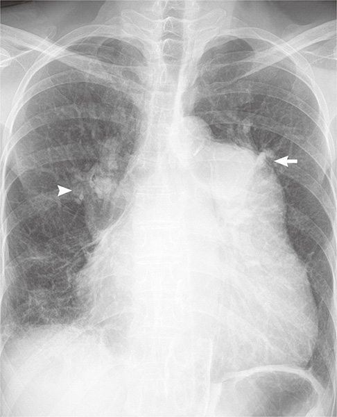
Fig. 1 Chest X-ray. Cardiomegaly involving right chambers, enlarged pulmonary trunk (arrow), and right main pulmonary artery (head arrow).
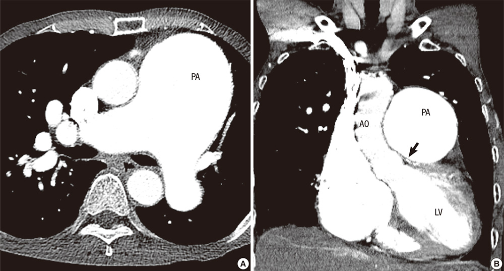
Fig. 2 Chest CT scan with contrast. (A) Markedly dilated pulmonary trunk (68mm) and pulmonary arteries. (B) Dilated main pulmonary artery trunk pressing against left main coronary artery. Arrows point to compression. AO, aorta; PA, main pulmonary artery trunk; LV, Left ventricle.

Fig. 3 Transthoracic echocardiogram shows evidence of a dilated main pulmonary artery trunk pressing against left main coronary artery.
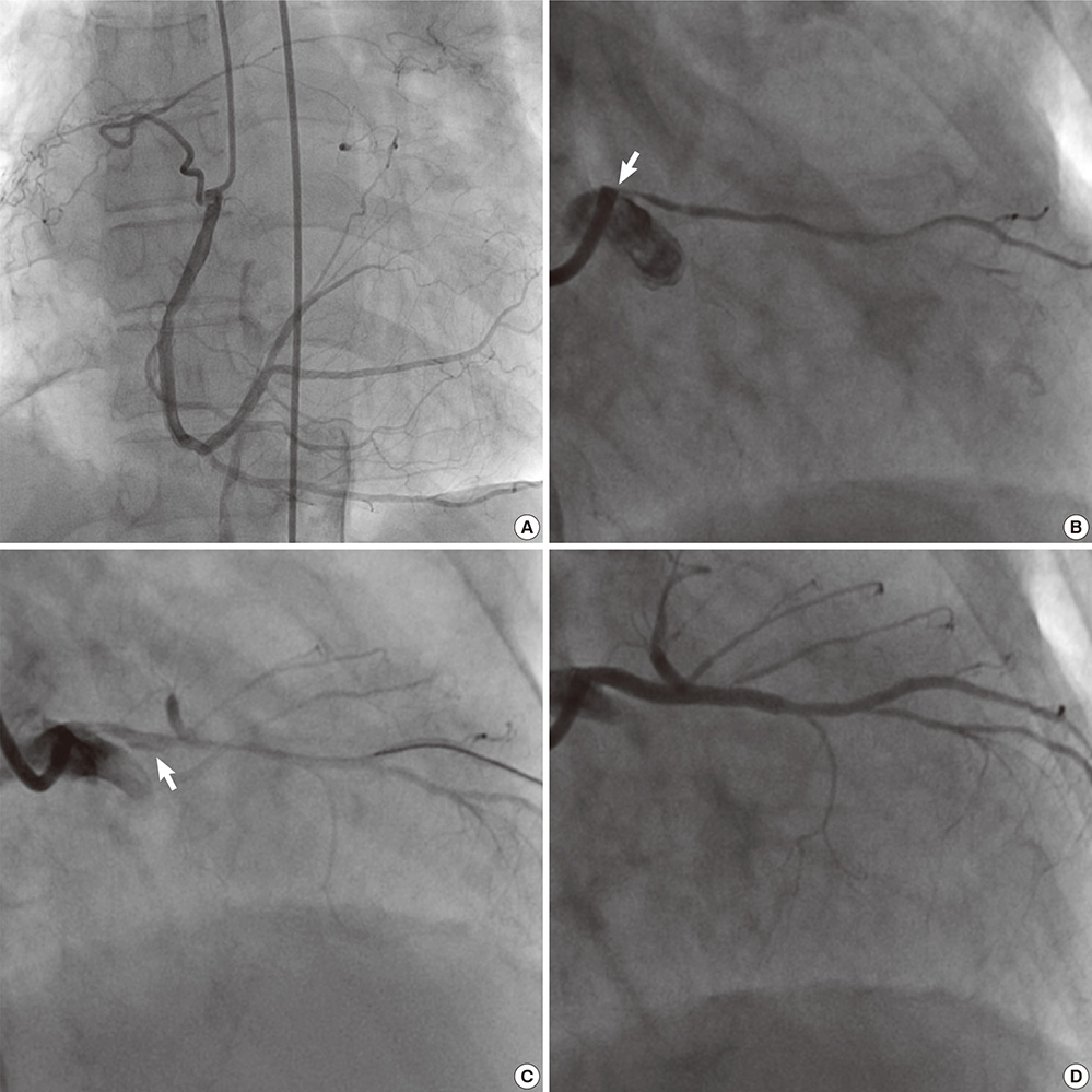
Fig. 4 Coronary angiography. (A) Right coronary angiography demonstrated minimal stenosis with grade 2 collaterals to the left circumflex artery and an arteriovenous fistula connecting with pulmonary artery. (B) Left coronary angiography demonstrated hypoplasia of left coronary artery and only diagonal branch was observed without left anterior descending (LAD) and left circumflex coronary artery (LCX). The study also reveals narrowing of the LMCA at its take-off from the aorta (arrow). (C) After balloon angioplasty, LAD and LCX was observed with LMCA dissection (arrow). (D) After stenting of the left main stenosis, excellent results with wide lumen of the left main coronary artery.
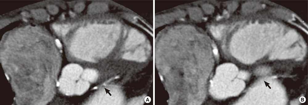
Fig. 5 ECG-gated, 128-slice multidetector computed tomography (MDCT) coronary angiography demonstrated extrinsic compression of the LMCA by dilated pulmonary arterial trunk (arrow).
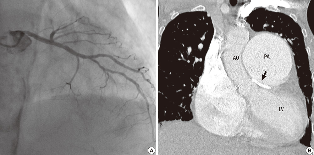
Fig. 6 One year after stenting follow-up. Coronary angiography (A) and a computed tomography with contrast (B) showed a widely patent stent in the LMCA in proximity of the dilated main pulmonary artery.
Reference
-
1. Rich S, Dantzker DR, Ayres SM, Bergofsky EH, Brundage BH, Detre KM, Fishman AP, Goldring RM, Groves BM, Koerner SK, et al. Primary pulmonary hypertension: a national prospective study. Ann Intern Med. 1987; 107:216–223.2. Vizza CD, Lynch JP, Ochoa LL, Richardson G, Trulock EP. Right and left ventricular dysfunction in patients with severe pulmonary disease. Chest. 1998; 113:576–583.3. D'Alonzo GE, Barst RJ, Ayres SM, Bergofsky EH, Brundage BH, Detre KM, Fishman AP, Goldring RM, Groves BM, Kernis JT, et al. Survival in patients with primary pulmonary hypertension: results from a national prospective registry. Ann Intern Med. 1991; 115:343–349.4. Kumar GV, Agarwal NB, Javali S, Patwardhan AM. Takayasu's arteritis with ostial and left main coronary artery stenosis. Tex Heart Inst J. 2007; 34:470–474.5. Rubín JM, Arias JC, Lambert JL. Unusual case of coronary stenosis caused by an external compression. Int J Cardiol. 1997; 62:167–169.6. Lee MS, Oyama J, Bhatia R, Kim YH, Park SJ. Left main coronary artery compression from pulmonary artery enlargement due to pulmonary hypertension: a contemporary review and argument for percutaneous revascularization. Catheter Cardiovasc Interv. 2010; 76:543–550.7. Kajita LJ, Martinez EE, Ambrose JA, Lemos PA, Esteves A, Nogueira da Gama M, Jatene AD, Ramires JA. Extrinsic compression of the left main coronary artery by a dilated pulmonary artery: clinical, angiographic, and hemodynamic determinants. Catheter Cardiovasc Interv. 2001; 52:49–54.8. Sivakumar K, Rajan M, Francis G, Murali K, Bashi V. Extrinsic compression of the left coronary ostium by the pulmonary trunk: management in a case of Eisenmenger syndrome. Tex Heart Inst J. 2010; 37:95–98.9. Lindsey JB, Brilakis ES, Banerjee S. Acute coronary syndrome due to extrinsic compression of the left main coronary artery in a patient with severe pulmonary hypertension: successful treatment with percutaneous coronary intervention. Cardiovasc Revasc Med. 2008; 9:47–51.10. Kawut SM, Silvestry FE, Ferrari VA, DeNofrio D, Axel L, Loh E, Palevsky HI. Extrinsic compression of the left main coronary artery by the pulmonary artery in patients with long-standing pulmonary hypertension. Am J Cardiol. 1999; 83:984–986.11. Schuijf JD, Jukema JW, van der Wall EE, Bax JJ. Multi-slice computed tomography in the evaluation of patients with acute chest pain. Acute Card Care. 2007; 9:214–221.12. De Jesus Perez VA, Haddad F, Vagelos RH, Fearon W, Feinstein J, Zamanian RT. Angina associated with left main coronary artery compression in pulmonary hypertension. J Heart Lung Transplant. 2009; 28:527–530.13. Rich S, McLaughlin VV, O'Neill W. Stenting to reverse left ventricular ischemia due to left main coronary artery compression in primary pulmonary hypertension. Chest. 2001; 120:1412–1415.14. Di Salvo G, Eyskens B, Claus P, D'hooge J, Bijnens B, Suys B, De Wolf D, Gewillig M, Sutherland GR, Mertens L. Late post-repair ventricular function in patients with origin of the left main coronary artery from the pulmonary trunk. Am J Cardiol. 2004; 93:506–508.15. Kushner FG, Hand M, Smith SC Jr, King SB 3rd, Anderson JL, Antman EM, Bailey SR, Bates ER, Blankenship JC, Casey DE Jr, et al. 2009 focused updates: ACC/AHA guidelines for the management of patients with ST-elevation myocardial infarction (updating the 2004 guideline and 2007 focused update) and ACC/AHA/SCAI guidelines on percutaneous coronary intervention (updating the 2005 guideline and 2007 focused update) a report of the American College of Cardiology Foundation/American Heart Association Task Force on Practice Guidelines. J Am Coll Cardiol. 2009; 54:2205–2241.16. Park SJ, Park SW, Hong MK, Cheong SS, Lee CW, Kim JJ, Hong MK, Mintz GS, Leon MB. Stenting of unprotected left main coronary artery stenoses: immediate and late outcomes. J Am Coll Cardiol. 1998; 31:37–42.17. Silvestri M, Barragan P, Sainsous J, Bayet G, Simeoni JB, Roquebert PO, Macaluso G, Bouvier JL, Comet B. Unprotected left main coronary artery stenting: immediate and medium-term outcomes of 140 elective procedures. J Am Coll Cardiol. 2000; 35:1543–1550.
- Full Text Links
-
- Actions
-
Cited
- CITED
-
- Close
- Share
-
- Similar articles
-
- Anomalous origin of the left coronary artery from the pulmonary artery
- Total Occlusion of Left Main Coronary Artery by Dilated Main Pulmonary Artery in a Patient with Severe Pulmonary Hypertension
- Congenital Absence of Left Circumflex Coronary Artery: Circumflex Artery Extended from Right Coronary Artery
- A Case of Percutaneous Transcatheter Coil Embolization for Congenital Coronary Arteriovenous Fistula
- A Case of Left Coronary Artery Milking Treated by Direct Stenting During Percutaneous Coronary Intervention in A Patient with Unstable Angina
