A Case of Castleman's Disease Mimicking a Hepatocellular Carcinoma: A Case Report and Review of Literature
- Affiliations
-
- 1Department of Internal Medicine and Hepatology Center, Bundang Jesaeng General Hospital, Seongnam, Korea. bohkim@medimail.co.kr
- 2Department of Radiology, Bundang Jesaeng General Hospital, Seongnam, Korea.
- 3Department of General Surgery, Bundang Jesaeng General Hospital, Seongnam, Korea.
- 4Department of Pathology, Bundang Jesaeng General Hospital, Seongnam, Korea.
- KMID: 1775834
- DOI: http://doi.org/10.4166/kjg.2012.59.1.53
Abstract
- Castleman's disease is a rare disease characterized by lymph node hyperplasia. Although Castleman's disease can occur wherever lymphoid tissue is found, it rarely appears in the abdominal cavity, and is especially rare adjacent to the liver. Here, we report a rare case of Castleman's disease in the portal area that mimicked a hepatocellular carcinoma (HCC) in a chronic hepatitis B patient. A 40 year-old woman with chronic hepatitis B presented with right upper quadrant discomfort. Computed tomography and magnetic resonance imaging results showed a 2.2 cm-sized, exophytic hypervascular mass in the portal area. HCC was suspected. However, histologic examination revealed Castleman's disease. We suggest that Castleman's disease should be included as a rare differential diagnosis of a hypervascular mass in the portal area, even in patients with chronic hepatitis B.
Keyword
MeSH Terms
-
Adult
Carcinoma, Hepatocellular/diagnosis
Diagnosis, Differential
Female
Giant Lymph Node Hyperplasia/complications/*diagnosis/pathology
Hepatitis B, Chronic/complications/diagnosis
Humans
Immunohistochemistry
Liver Neoplasms/diagnosis
Magnetic Resonance Imaging
Receptors, Complement 3d/metabolism
Tomography, X-Ray Computed
Figure
-
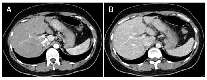
Fig. 1 Liver CT findings. (A) CT image on hepatic arterial phase showed a 2.1 cm diameter enhancing mass in the caudate lobe of the liver (arrow). (B) CT image on delayed phase showed that the nodule became isodense, compared with the liver parenchyma.
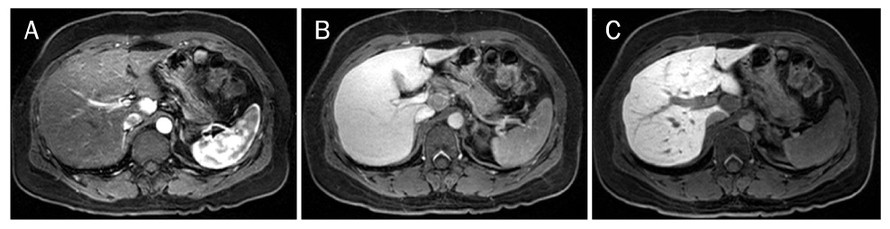
Fig. 2 Liver MRI findings. (A) Contrast-enhanced (gadolinium ethoxybenzyl diethylenetriamine pentaacetic acid) T1-weighted MRI on hepatic arterial phase showed a 2.2 cm diameter, exophytic enhancing nodular lesion in the caudate lobe of the liver. (B, C) Contrast-enhanced T1-weighted MRI on delayed phase (at 3 minutes and 20 minutes) showed no contrast uptake in the hepatic nodule.
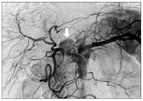
Fig. 3 Angiography findings. Celiac angiography showed a round extrahepatic hypervascular mass supplied by the common hepatic artery (arrow).
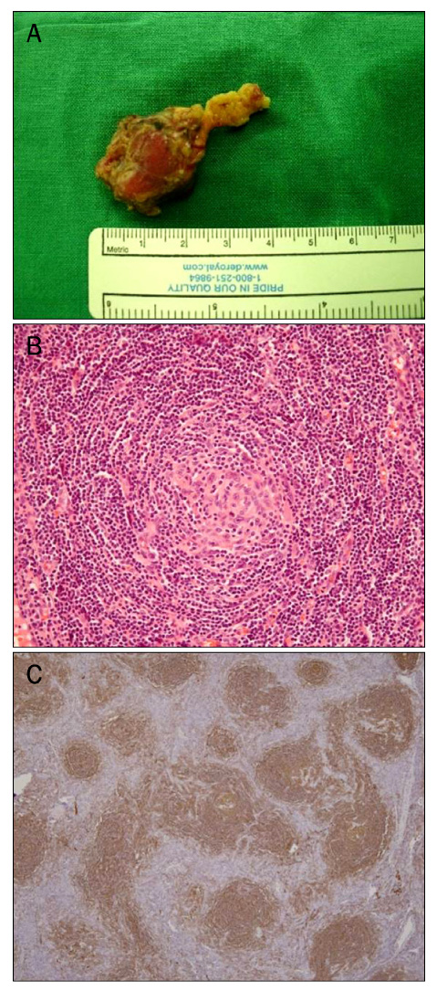
Fig. 4 Gross and histologic findings. (A) Photograph of the surgical specimen revealed a round mass with encapsulation. (B) Regressed germinal center replaced by hyalinized vessels and surrounded by concentrically arranged small lymphocytes (H&E, ×200). (C) Immunohistochemical stains for CD21 showing an expanded network of multiple tight collections of follicular dendritic cells (CD21, ×40).
Reference
-
1. Castleman B, Iverson L, Menendes VP. Localized mediastinal lymph-node hyperplasia resembling thymoma. Cancer. 1956. 9:822–830.2. Keller AR, Hochholzer L, Castleman B. Hyaline-vascular and plasma-cell types of giant lymph node hyperplasia of the mediastinum and other locations. Cancer. 1972. 29:670–683.3. Gaba AR, Stein RS, Sweet DL, Variakojis D. Multicentric giant lymph node hyperplasia. Am J Clin Pathol. 1978. 69:86–90.4. Casper C. The aetiology and management of Castleman disease at 50 years: translating pathophysiology to patient care. Br J Haematol. 2005. 129:3–17.5. van Rhee F, Stone K, Szmania S, Barlogie B, Singh Z. Castleman disease in the 21st century: an update on diagnosis, assessment, and therapy. Clin Adv Hematol Oncol. 2010. 8:486–498.6. Bowne WB, Lewis JJ, Filippa DA, et al. The management of unicentric and multicentric Castleman's disease: a report of 16 cases and a review of the literature. Cancer. 1999. 85:706–717.7. Bucher P, Chassot G, Zufferey G, Ris F, Huber O, Morel P. Surgical management of abdominal and retroperitoneal Castleman's disease. World J Surg Oncol. 2005. 3:33.8. Madan R, Chen JH, Trotman-Dickenson B, Jacobson F, Hunsaker A. The spectrum of Castleman's disease: Mimics, radiologic pathologic correlation and role of imaging in patient management. Eur J Radiol. 2012. 81:123–131.9. Rahmouni A, Golli M, Mathieu D, Anglade MC, Charlotte F, Vasile N. Castleman disease mimicking liver tumor: CT and MR features. J Comput Assist Tomogr. 1992. 16:699–703.10. Farkas E, Tóth B, Besznyák I. Retroperitoneal Castleman tumor. Orv Hetil. 1993. 134:413–416.11. Peck D, Lum PA. Castleman disease in the porta hepatis: biphasic helical computed tomography. Can Assoc Radiol J. 1996. 47:410–412.12. Cirillo RL Jr, Vitellas KM, Deyoung BR, Bennett WF. Castleman disease mimicking a hepatic neoplasm. Clin Imaging. 1998. 22:124–129.13. Uzunlar AK, Ozateş M, Yaldiz M, Büyükbayram H, Ozaydin M. Castleman's disease in the porta hepatis. Eur Radiol. 2000. 10:1913–1915.14. Al-Salamah SM, Khan IA, Khalid K, Al-Shakweer WA. Castleman disease presenting as obstructive jaundice. Saudi Med J. 2005. 26:111–113.15. Sato N, Kondo S, Saito K, et al. Hyaline vascular-type Castleman's disease in the hepatoduodenal ligament: report of a case. Surg Today. 2006. 36:647–650.16. Karami H, Sahebpour AA, Ghasemi M, et al. Hyaline vascular-type Castleman's disease in the hilum of liver: a case report. Cases J. 2010. 3:74.17. User IR, Akçören Z, Karnak I. Castleman disease: an unusual diagnosis of a portal mass in an 8-year-old boy. J Pediatr Surg. 2011. 46:e9–e11.18. Waterston A, Bower M. Fifty years of multicentric Castleman's disease. Acta Oncol. 2004. 43:698–704.19. Chung YE, Kim MJ, Park YN, et al. Varying appearances of cholangiocarcinoma: radiologic-pathologic correlation. Radiographics. 2009. 29:683–700.20. Chronowski GM, Ha CS, Wilder RB, Cabanillas F, Manning J, Cox JD. Treatment of unicentric and multicentric Castleman disease and the role of radiotherapy. Cancer. 2001. 92:670–676.
- Full Text Links
-
- Actions
-
Cited
- CITED
-
- Close
- Share
-
- Similar articles
-
- Castleman Disease in the Kidney and Retroperitoneum Mimicking Renal Cell Carcinoma with Retroperitoneal Lymphadenopathy: A Case Report
- A Case of Localized Castleman's Disease in a Patient with Rheumatoid Arthritis
- Intrahepatic Splenosis Mimicking Hepatocellular Carcinoma: A Case Report
- Castleman Disease Arising from IVlesentery: A Case Report
- Cutaneous Metastasis from Hepatocellular Carcinoma Mimicking Pyogenic Granuloma
