A Case of Spindle Cell Carcinoma of the Stomach Presenting with Hematochezia and Weight Loss Due to Fistulous Tract Formation with Colon
- Affiliations
-
- 1Department of Internal Medicine, The Catholic University of Korea College of Medicine, Seoul, Korea. adagio@catholic.ac.kr
- 2Department of Surgery, The Catholic University of Korea College of Medicine, Seoul, Korea.
- 3Department of Pathology, The Catholic University of Korea College of Medicine, Seoul, Korea.
- KMID: 1775751
- DOI: http://doi.org/10.4166/kjg.2013.62.2.126
Abstract
- Spindle cell carcinoma (SpCC) is a rare tumor consisting of spindle cells which express cytokeratin. Despite recent advances in immunohistochemical and genetic studies, precise histogenesis of SpCC is still controversial and this tumor had been referred to with a wide range of names (in the past): carcinosarcoma, pseudosarcoma, sarcomatoid carcinoma, pseudosarcomatous carcinoma, and collision tumor. Recently, the authors experienced an extremely rare case of SpCC arising from the stomach. A 64-year-old male presented with unintended weight loss and hematochezia. Endoscopic examination revealed a fistulous tract between the stomach and the transverse colon which was made by direct invasion of SpCC of the stomach to the colon. Histologically, the tumor was positive for both vimentin and cytokeratin but negative for CD117, CD34, actin, and desmin. Herein, we report a case of SpCC arising from the stomach that formed a fistulous tract with the colon which was diagnosed during evaluation of hematochezia and weight loss.
Keyword
MeSH Terms
-
Antineoplastic Agents/therapeutic use
Brain Neoplasms/secondary
Carcinoma/*diagnosis/drug therapy/pathology
Colon, Transverse
Endoscopy, Digestive System
Fistula/pathology
Gastrointestinal Hemorrhage/etiology
Humans
Keratins/metabolism
Magnetic Resonance Imaging
Male
Middle Aged
Stomach Neoplasms/*diagnosis/drug therapy/pathology
Tomography, X-Ray Computed
Weight Loss
Antineoplastic Agents
Keratins
Figure
-

Fig. 1. Endoscopic findings of spindle cell carcinoma. (A) Esophagogastroduodenoscopy shows a large ulceroinfiltrative lesion with fistulous opening on the greater curvature side of the gastric body. (B) Colonoscopy also shows ulceroinfiltrative lesion on the mid transverse colon.

Fig. 2. Histological findings of spindle cell carcinoma. (A) Diffuse proliferation of spindle-shaped cells with infiltration of neutrophils is seen (H&E,×40). (B) Atypical spindle-shaped cells with nuclear enlargement and polymorphism are observed (H&E, ×400). Both vimentin (C) and cytokeratin (D) are positively stained on the same section (immunohistochemical stain, ×100).
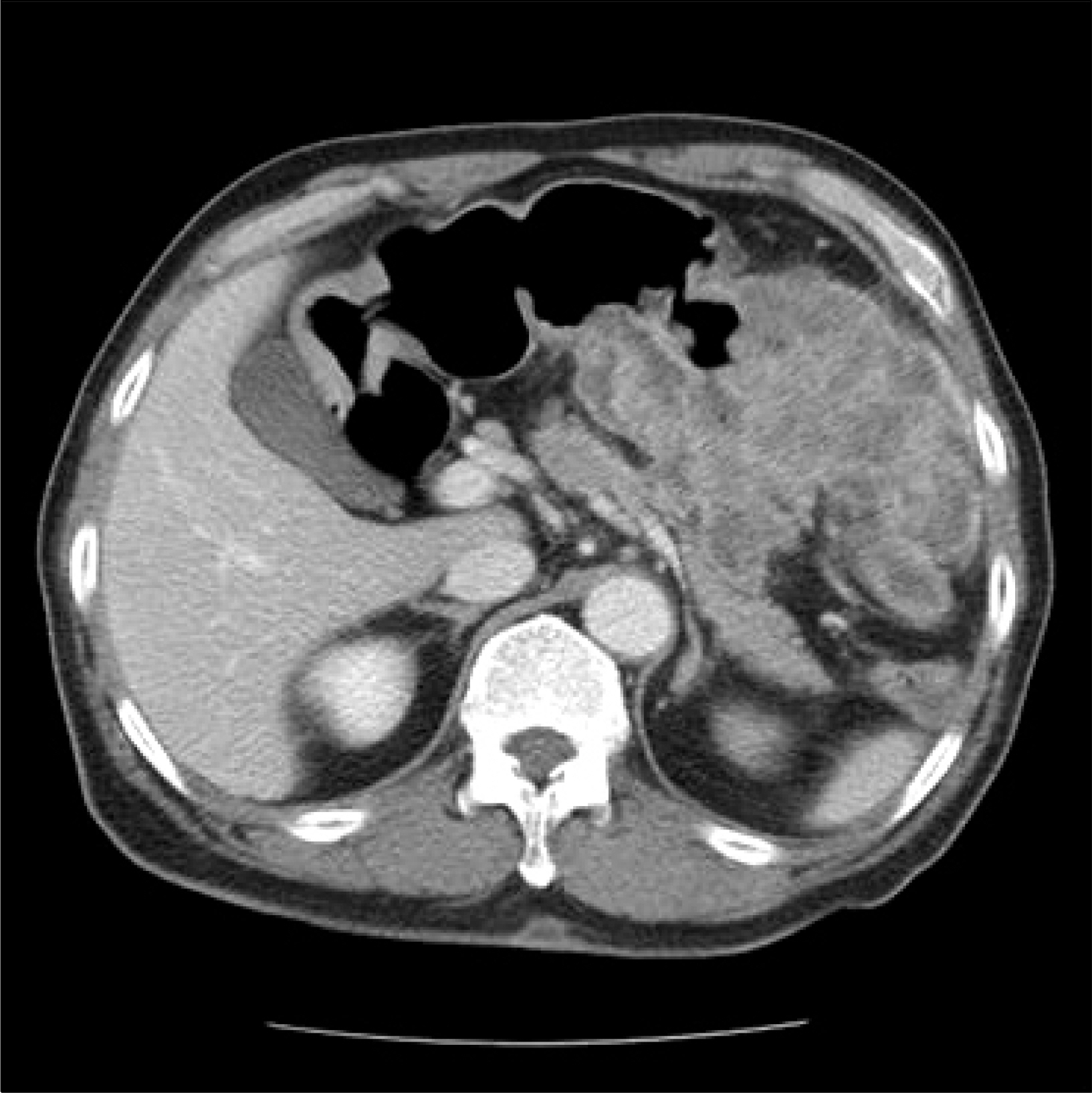
Fig. 3. Abdominal CT scan shows irregular wall thickening of the greater curvature side of gastric body with transverse colon invasion.
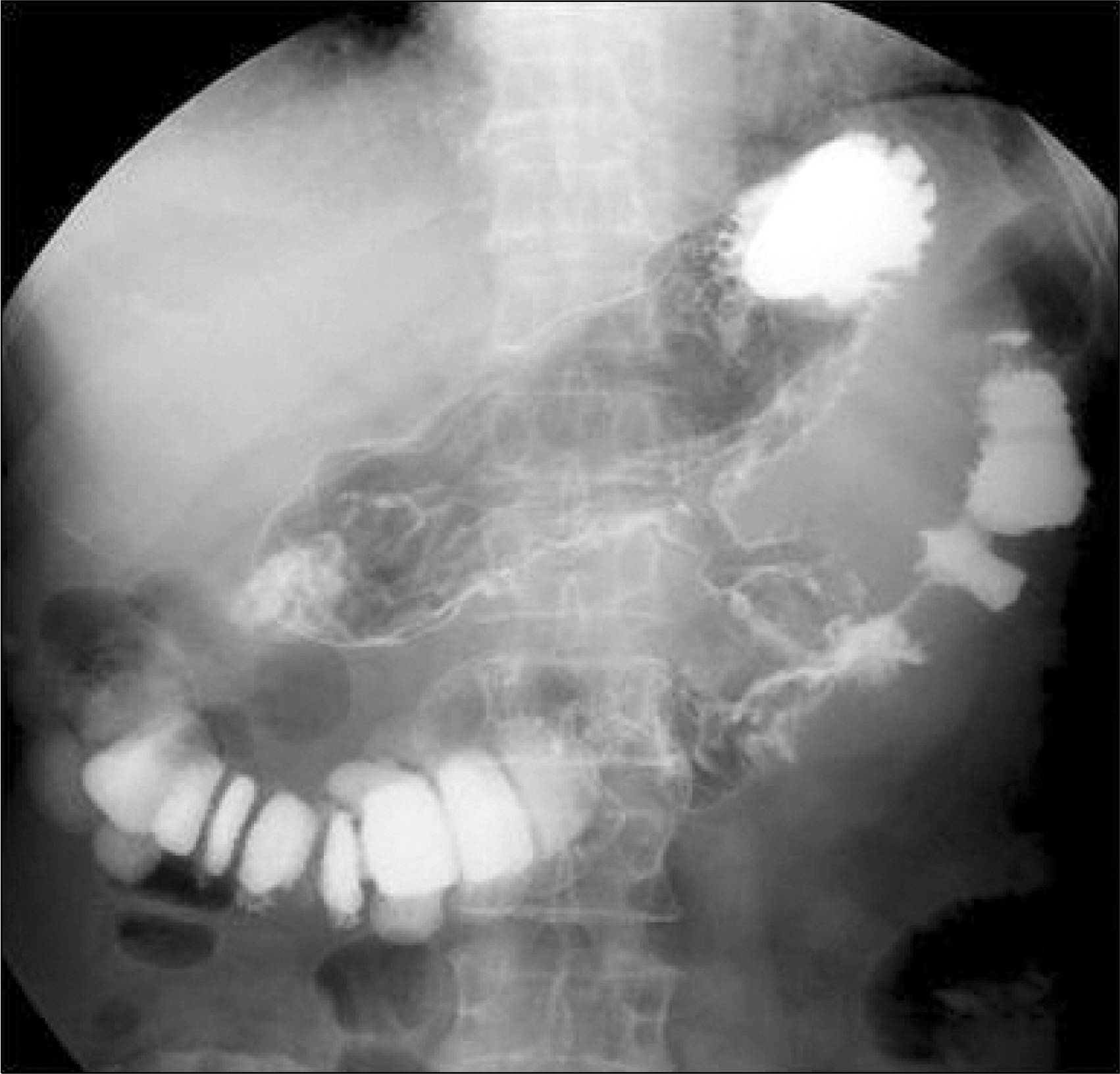
Fig. 4. Upper gastrointestinal series shows fistula between the gastric body and transverse colon without peritoneal leakage.
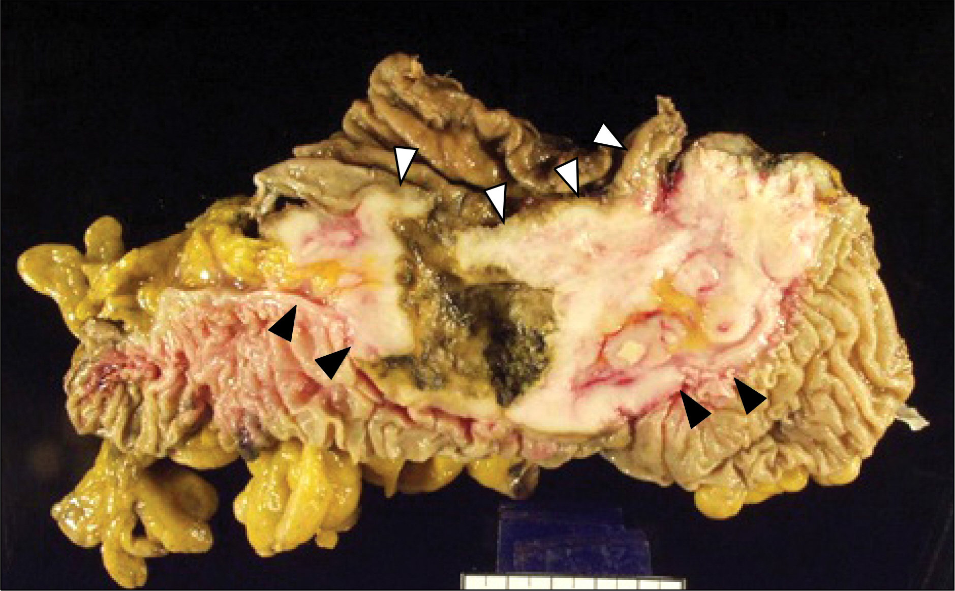
Fig. 5. Macroscopic features of the specimen. Gastric wall is thickened due to infiltration by spindle cell carcinoma (white arrowheads). The mass invades directly to adjacent fat tissue and transverse colon (black arrowheads). The central portion of the mass underwent necrotic change and forms a fistulous tract between the stomach and colon.
Reference
-
References
1. Taniyama K, Sasaki N, Mukai T, et al. Carcinosarcomas of the esophagus. Pathol Int. 1995; 45:297–302.
Article2. Gal AA, Martin SE, Kernen JA, Patterson MJ. Esophageal carcinoma with prominent spindle cells. Cancer. 1987; 60:2244–2250.
Article3. Iyomasa S, Kato H, Tachimori Y, Watanabe H, Yamaguchi H, Itabashi M. Carcinosarcoma of the esophagus: a twenty-case study. Jpn J Clin Oncol. 1990; 20:99–106.4. Kayaselcuk F, Tuncer I, Toyganözü Y, Bal N, Ozgür G. Carcinosarcoma of the stomach. Pathol Oncol Res. 2002; 8:275–277.5. Oak JH, Chung WC, Jung JH, et al. A case of carcinosarcoma in a patient with corrosive esophagitis. Korean J Gastroenterol. 2008; 52:42–47.6. Kim JC, Lee JM, Jung SE, Lee KY, Hahn ST, Kim MD. Spindle-cell carcinoma of esophagus: a case report. J Korean Radiol Soc. 2001; 44:593–597.
Article7. Kim YH, Kim YK, Lee MG, et al. A case of spindle cell carcinoma of the esophagus. Korean J Gastrointest Endosc. 1998; 18:691–697.8. Maiorana A, Fante R, Maria Cesinaro A, Adriana Fano R. Synchronous occurrence of epithelial and stromal tumors in the stomach: a report of 6 cases. Arch Pathol Lab Med. 2000; 124:682–686.9. Randjelovic T, Filipovic B, Babic D, Cemerikic V, Filipovic B. Carcinosarcoma of the stomach: a case report and review of the literature. World J Gastroenterol. 2007; 13:5533–5536.
Article10. Teramachi K, Kanomata N, Hasebe T, Ishii G, Sugito M, Ochiai A. Carcinosarcoma (pure endocrine cell carcinoma with sarcoma components) of the stomach. Pathol Int. 2003; 53:552–556.
Article11. Matsui K, Jin XM, Kitagawa M, Miwa A. Clinicopathologic features of neuroendocrine carcinomas of the stomach: appraisal of small cell and large cell variants. Arch Pathol Lab Med. 1998; 122:1010–1017.12. Sato Y, Shimozono T, Kawano S, et al. Gastric carcinosarcoma, coexistence of adenosquamous carcinoma and rhabdomyosarcoma: a case report. Histopathology. 2001; 39:543–544.
Article
- Full Text Links
-
- Actions
-
Cited
- CITED
-
- Close
- Share
-
- Similar articles
-
- Spindle Cell Carcinoma in Larynx: A case showing bone formation
- An Unusual Cause of Gastrointestinal Hemorrhage: Gastrocolic Fistula Caused by Colon Cancer Invasion
- A Case of Malignant Coloenteric Fistula
- A Case of Spindle Cell Carcinoma Accompanied by Inverted Papilloma
- A Case of Primary Squamous Cell Carcinoma of Sigmoid Colon
