Ann Pediatr Endocrinol Metab.
2014 Dec;19(4):202-207. 10.6065/apem.2014.19.4.202.
Clinical and radiological features of pituitary stalk lesions in children and adolescents
- Affiliations
-
- 1Department of Pediatrics, Seoul National University Children's Hospital, Seoul National University College of Medicine, Seoul, Korea.
- 2Department of Pediatrics, SMG-SNU Boramae Medical Center, Seoul, Korea. gnoygnoes@hanmail.net
- KMID: 1769353
- DOI: http://doi.org/10.6065/apem.2014.19.4.202
Abstract
- PURPOSE
The diagnosis of pituitary stalk lesion has been based on clinical feature, radiologic assessment for its critical location and role. This study aimed to investigate clinical symptoms, endocrine disturbance, magnetic resonance imaging (MRI) findings of pituitary stalk lesions in children and adolescents and to evaluate differences between neoplastic lesions with the others.
METHODS
We performed a retrospective review of patients under 18 years old with pituitary stalk lesions diagnosed at the Seoul National University Children's Hospital between 2000 and 2013, by a text search for head MRI reports by using 'pituitary stalk', 'infundibulum', and 'infundibular stalk', as keywords.
RESULTS
For the 76 patients, sixteen patients (21.1%) had congenital lesions, and 52 (68.4%) had neoplasms. No inflammatory lesions were found. Diabetes insipidus (DI) was the most common endocrine defect, diagnosed in 38 patients (50%). There was male predominance especially in neoplastic group. Thickened pituitary stalk was, but enhancement of lesion was not, associated with neoplasm. DI was more prevalent in neoplastic stalk lesions. Anterior pituitary dysfunction such as growth hormone and adrenocorticotropic hormone deficiencies were less prevalent in neoplastic lesions of pituitary stalk.
CONCLUSION
In conclusion, the etiology of pituitary stalk lesions in children and adolescents is diverse and different from that in adults. Neoplastic pituitary stalk lesions can be differentiated from nonneoplastic lesions by systemic evaluation of clinical, hormonal, radiological findings.
Keyword
MeSH Terms
Figure
-
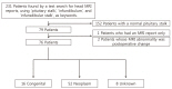
Fig. 1 A total 231 patients was found whose magnetic resonance imaging (MRI) reports included key words such as 'pituitary stalk', 'infundibulum', and 'infundibular stalk'. One hundred and fifty two patients with a normal pituitary stalk in their MRI report, 1 patient who had an MRI report only, and 2 patients with postoperative change, were excluded. Of the remaining 76 patients, 16 patients had congenital lesions, 52 patients neoplasm, and 8 patients unknown etiologies.
Reference
-
1. Simmons GE, Suchnicki JE, Rak KM, Damiano TR. MR imaging of the pituitary stalk: size, shape, and enhancement pattern. AJR Am J Roentgenol. 1992; 159:375–377. PMID: 1632360.2. Rupp D, Molitch M. Pituitary stalk lesions. Curr Opin Endocrinol Diabetes Obes. 2008; 15:339–345. PMID: 18594274.3. Turcu AF, Erickson BJ, Lin E, Guadalix S, Schwartz K, Scheithauer BW, et al. Pituitary stalk lesions: the Mayo Clinic experience. J Clin Endocrinol Metab. 2013; 98:1812–1818. PMID: 23533231.4. Hamilton BE, Salzman KL, Osborn AG. Anatomic and pathologic spectrum of pituitary infundibulum lesions. AJR Am J Roentgenol. 2007; 188:W223–W232. PMID: 17312027.5. Marchand I, Barkaoui MA, Garel C, Polak M, Donadieu J. Writing Committee. Central diabetes insipidus as the inaugural manifestation of Langerhans cell histiocytosis: natural history and medical evaluation of 26 children and adolescents. J Clin Endocrinol Metab. 2011; 96:E1352–E1360. PMID: 21752883.
Article6. Di Iorgi N, Allegri AE, Napoli F, Calcagno A, Calandra E, Fratangeli N, et al. Central diabetes insipidus in children and young adults: etiological diagnosis and long-term outcome of idiopathic cases. J Clin Endocrinol Metab. 2014; 99:1264–1272. PMID: 24276447.
Article7. Beni-Adani L, Sainte-Rose C, Zerah M, Brunelle F, Constantini S, Renier D, et al. Surgical implications of the thickened pituitary stalk accompanied by central diabetes insipidus. J Neurosurg. 2005; 103(2 Suppl):142–147. PMID: 16370280.
Article8. Bihan H, Christozova V, Dumas JL, Jomaa R, Valeyre D, Tazi A, et al. Sarcoidosis: clinical, hormonal, and magnetic resonance imaging (MRI) manifestations of hypothalamicpituitary disease in 9 patients and review of the literature. Medicine (Baltimore). 2007; 86:259–268. PMID: 17873755.9. Twilt M, Benseler SM. Childhood inflammator y brain diseases: pathogenesis, diagnosis and therapy. Rheumatology (Oxford). 2014; 53:1359–1368. PMID: 24324213.
Article10. Nozaki K, Scott TF, Sohn M, Judson MA. Isolated neurosarcoidosis: case series in 2 sarcoidosis centers. Neurologist. 2012; 18:373–377. PMID: 23114669.11. Oksanen V. Neurosarcoidosis: clinical presentations and course in 50 patients. Acta Neurol Scand. 1986; 73:283–290. PMID: 3716768.
Article12. Iannuzzi MC, Rybicki BA, Teirstein AS. Sarcoidosis. N Engl J Med. 2007; 357:2153–2165. PMID: 18032765.
Article13. Pietinalho A, Hiraga Y, Hosoda Y, Lofroos AB, Yamaguchi M, Selroos O. The frequency of sarcoidosis in Finland and Hokkaido, Japan. A comparative epidemiological study. Sarcoidosis. 1995; 12:61–67. PMID: 7617979.14. Scholten V, ten Hove WM, Macdonald EA. An unusual presentation of neurosarcoidosis in an 11-year-old boy. Can J Neurol Sci. 2009; 36:783–786. PMID: 19960762.
Article
- Full Text Links
-
- Actions
-
Cited
- CITED
-
- Close
- Share
-
- Similar articles
-
- Sporadic Hemangioblastoma in the Pituitary Stalk: A Case Report and Review of the Literature
- Surgical Treatment of Hemangioblastoma in the Pituitary Stalk: An Extremely Rare Case
- Pituitary Stalk Hemangioblastoma in a von Hippel-Lindau Patient : Clinical Course Follow-Up Over a 20-Year Period
- Pituitary Stalk Transection Syndrome
- Suprasellar Mass Lesions Presenting with Central Diabets Insipidus
