Pigment Deposition of Cosmetic Contact Lenses on the Cornea after Intense Pulsed-Light Treatment
- Affiliations
-
- 1HanGil Eye Hospital, Incheon, Korea. limthnet@naver.com
- KMID: 1769092
- DOI: http://doi.org/10.3341/kjo.2010.24.6.367
Abstract
- We report a case of corneal deposition of pigments from cosmetic contact lenses after intense pulsed-light (IPL) therapy. A 30-year-old female visited our outpatient clinic with ocular pain and epiphora in both eyes; these symptoms developed soon after she had undergone facial IPL treatment. She was wearing cosmetic contact lenses throughout the IPL procedure. At presentation, her uncorrected visual acuity was 2/20 in both eyes, and the slit-lamp examination revealed deposition of the color pigment of the cosmetic contact lens onto the corneal epithelium. We scraped the corneal epithelium along with the deposited pigments using a no. 15 blade; seven days after the procedure, the corneal epithelium had healed without any complications. This case highlights the importance of considering the possibility of ocular complications during IPL treatment, particularly in individuals using contact lenses. To prevent ocular damage, IPL procedures should be performed only after removing the lenses and applying eyeshields.
MeSH Terms
Figure
-
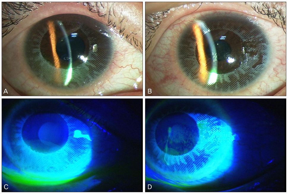
Fig. 1 Slit-lamp photographs at the first examination (A,C, right eye; B,D, left eye). (A,B) Conjunctival injection, mild chemosis and deposition of the color pigments of the cosmetic contact lens onto the corneal epithelium were observed. The pattern of the deposition matched the pattern on the contact lenses. (C,D) Fluorescein-dye staining revealed punctate epithelial erosions and corneal epithelial defects in both eyes.

Fig. 2 Photographs taken immediately after the excision of the corneal epithelium with the no. 15 blade. The pigments were completely excised. Therapeutic lenses were implanted. (A) Right eye. (B) Left eye.
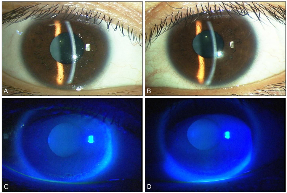
Fig. 3 Slit-lamp photographs taken on the 7th day after treatment (A,C, right eye; B,D, left eye). (A,B) Corneal epithelial defects and erosions were completely healed. The cornea was clear, and the anterior chamber was clear. (C,D) These are the same photographs after fluorescein staining. An intact epithelium was observed.
Reference
-
1. Phillips AJ, Speedwell L, editors. Contact lenses. 2007. 5th ed. London: Butterworth Heinemann;519–530.2. Lim TH, Lee JR, Choi KY, et al. Corneal melting and descemetocele resulting from noninfectious keratitis related to the cosmetic contact lenses. J Korean Ophthalmol Soc. 2009. 50:774–778.3. Steinemann TL, Pinninti U, Szczotka LB, et al. Ocular complications associated with the use of cosmetic contact lenses from unlicensed vendors. Eye Contact Lens. 2003. 29:196–200.4. Steinemann TL, Fletcher M, Bonny AE, et al. Over-the-counter decorative contact lenses: cosmetic or medical devices? A case series. Eye Contact Lens. 2005. 31:194–200.5. Raulin C, Greve B, Grema H. IPL technology: a review. Lasers Surg Med. 2003. 32:78–87.6. Goldberg DJ, Cutler KB. Nonablative treatment of rhytids with intense pulsed light. Lasers Surg Med. 2000. 26:196–200.7. Bitter PH. Noninvasive rejuvenation of photodamaged skin using serial, full-face intense pulsed light treatments. Dermatol Surg. 2000. 26:835–842.8. Gagnon MR, Walter KA. A case of acanthamoeba keratitis as a result of a cosmetic contact lens. Eye Contact Lens. 2006. 32:37–38.9. Carkeet A. Field restriction and vignetting in contact lenses with opaque peripheries. Clin Exp Optom. 1998. 81:151–158.10. Hiraoka T, Ishii Y, Okamoto F, Oshika T. Influence of cosmetically tinted soft contact lenses on higher-order wavefront aberrations and visual performance. Graefes Arch Clin Exp Ophthalmol. 2009. 247:225–233.11. Choi HW, Moon SW, Nam KH, Chung SH. Late-onset interface inflammation associated with wearing cosmetic lenses 18 months after laser in situ keratomileusis. Cornea. 2008. 27:252–254.
- Full Text Links
-
- Actions
-
Cited
- CITED
-
- Close
- Share
-
- Similar articles
-
- Characteristics and Complications of Cosmetic Contact Lens
- Corneal Melting and Descemetocele Resulting From Noninfectious Keratitis Related to the Cosmetic Contact Lenses
- Two Cases of Poikiloderma of Neck and Chest Treated by Intense Pulsed Light
- Physiological Changes in the Cornea When Wearing Rigid Gas Permeable Contact Lenses
- Characteristics of Scleral Lenses and Patient Selection
