Korean J Ophthalmol.
2010 Dec;24(6):360-363. 10.3341/kjo.2010.24.6.360.
Brain Imaging Studies in Leber's Congenital Amaurosis: New Radiologic Findings Associated with the Complex Trait
- Affiliations
-
- 1Department of Ophthalmology, Seoul National University College of Medicine, Seoul, Korea. ysyu@snu.ac.kr
- 2Department of Ophthalmology, Seoul National University Bundang Hospital, Seongnam, Korea.
- 3Department of Laboratory Medicine, Seoul National University College of Medicine, Seoul, Korea.
- 4Seoul Artificial Eye Center, Seoul National University Hospital Clinical Research Institute, Seoul, Korea.
- KMID: 1769090
- DOI: http://doi.org/10.3341/kjo.2010.24.6.360
Abstract
- PURPOSE
To report the incidence and new findings of abnormal brain imaging studies associated with patients initially diagnosed with Leber's congenital amaurosis (LCA) without definite systemic abnormalities and to determine the need for brain imaging studies in these patients.
METHODS
A retrospective review of medical records was performed in 83 patients initially diagnosed as LCA and without definite systemic abnormalities before the age of 6 months in 2 tertiary referral centers. Brain magnetic resonance imaging was performed in 31 of 83 patients (37.3%).
RESULTS
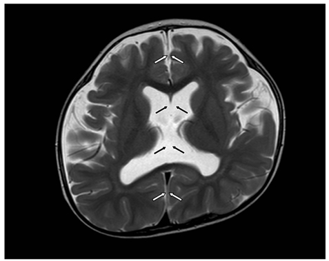
Six of 31 patients (19%) had radiologically documented brain abnormalities. Two patients had cerebellar vermis hypoplasia, 1 patient showed an absence of septum pellucidum, 2 subjects showed mild external hydrocephalus, and 1 patient was found to have a small cerebellum.
CONCLUSIONS
Approximately one fifth of the LCA patients in whom brain imaging was performed were associated with brain abnormalities, including the absence of septum pellucidum, which has not been documented in the literature. Brain imaging is mandatory in patients primarily diagnosed with LCA, even without definite neurologic or systemic abnormalities.
Keyword
MeSH Terms
Figure
Reference
-
1. De Laey JJ. Leber's congenital amaurosis. Bull Soc Belge Ophtalmol. 1991. 241:41–50.2. Schroeder R, Mets MB, Maumenee IH. Leber's congenital amaurosis. Retrospective review of 43 cases and a new fundus finding in two cases. Arch Ophthalmol. 1987. 105:356–359.3. Fazzi E, Signorini SG, Uggetti C, et al. Towards improved clinical characterization of Leber congenital amaurosis: neurological and systemic findings. Am J Med Genet A. 2005. 132A:13–19.4. Lambert SR, Kriss A, Taylor D, et al. Follow-up and diagnostic reappraisal of 75 patients with Leber's congenital amaurosis. Am J Ophthalmol. 1989. 107:624–631.5. Hwang JS, Kim JH, Choung HK, et al. Clinical characteristics of Leber's congenital amaurosis in Korea. J Korean Ophthalmol Soc. 2007. 48:1257–1262.6. Casteels I, Spileers W, Demaerel P, et al. Leber congenital amaurosis: differential diagnosis, ophthalmological and neuroradiological report of 18 patients. Neuropediatrics. 1996. 27:189–193.7. Steinberg A, Ronen S, Zlotogorski Z, et al. Central nervous system involvement in Leber congenital amaurosis. J Pediatr Ophthalmol Strabismus. 1992. 29:224–227.8. Weinstein JM, Gleaton M, Weidner WA, Young RS. Leber's congenital amaurosis. Relationship of structural CNS anomalies to psychomotor retardation. Arch Neurol. 1984. 41:204–206.9. Reynell J. Developmental patterns of visually handicapped children. Child Care Health Dev. 1978. 4:291–303.10. Waugh MC, Chong WK, Sonksen P. Neuroimaging in children with congenital disorders of the peripheral visual system. Dev Med Child Neurol. 1998. 40:812–819.11. Barkovich AJ, Norman D. Absence of the septum pellucidum: a useful sign in the diagnosis of congenital brain malformations. AJR Am J Roentgenol. 1989. 152:353–360.12. Yano S, Oda K, Watanabe Y, et al. Two sib cases of Leber congenital amaurosis with cerebellar vermis hypoplasia and multiple systemic anomalies. Am J Med Genet. 1998. 78:429–432.13. Morgan SA, Emsellem HA, Sandler JR. Absence of the septum pellucidum. Overlapping clinical syndromes. Arch Neurol. 1985. 42:769–770.14. Kuhn MJ, Swenson LC, Youssef HT. Absence of the septum pellucidum and related disorders. Comput Med Imaging Graph. 1993. 17:137–147.15. Williams J, Brodsky MC, Griebel M, et al. Septo-optic dysplasia: the clinical insignificance of an absent septum pellucidum. Dev Med Child Neurol. 1993. 35:490–501.16. Satran D, Pierpont ME, Dobyns WB. Cerebello-oculo-renal syndromes including Arima, Senior-Löken and COACH syndromes: more than just variants of Joubert syndrome. Am J Med Genet. 1999. 86:459–469.17. Gleeson JG, Keeler LC, Parisi MA, et al. Molar tooth sign of the midbrain-hindbrain junction: occurrence in multiple distinct syndromes. Am J Med Genet A. 2004. 125A:125–134.18. Chance PF, Cavalier L, Satran D, et al. Clinical nosologic and genetic aspects of Joubert and related syndromes. J Child Neurol. 1999. 14:660–666.19. Maria BL, Boltshauser E, Palmer SC, Tran TX. Clinical features and revised diagnostic criteria in Joubert syndrome. J Child Neurol. 1999. 14:583–590.20. Traboulsi EI, Koenekoop R, Stone EM. Lumpers or splitters? The role of molecular diagnosis in Leber congenital amaurosis. Ophthalmic Genet. 2006. 27:113–115.
- Full Text Links
- Actions
-
Cited
- CITED
-
- Close
- Share
- Similar articles
-
- Joubert Syndrome Associated with Leber's Congenital Amaurosis
- Clinical Characteristics of Leber's Congenital Amaurosis in Korea
- Two Cases of Senior-Loken Syndrome in Siblings
- Clinical features of Senior–Loken syndrome with IQCB1/NPHP5 mutation in a Filipino man
- Short-Term Outcomes of the First in Vivo Gene Therapy for RPE65-Mediated Retinitis Pigmentosa


