Korean J Gastroenterol.
2009 May;53(5):324-328. 10.4166/kjg.2009.53.5.324.
Pancreatic Tuberculosis Presenting with Pancreatic Cystic Tumor: A Case Report and Review of the Literature
- Affiliations
-
- 1Department of Internal Medicine, Chonnam National University Medical School, Gwangju, Korea. portalvein@naver.com
- KMID: 1767697
- DOI: http://doi.org/10.4166/kjg.2009.53.5.324
Abstract
- Pancreatic tuberculosis is a rare clinical entity, presenting as malignancy mimicking pancreatic mass. Therefore, it represents a diagnostic challenge. To date, ten cases have been reported in Korea. I report an additional case and review all Korean reports about pancreatic tuberculosis. A 57-year-old woman presented with abdominal pain. Abdominal computed tomography (CT) revealed a 2.2x1.2 cm cystic mass in pancreatic body. She was followed for nine months, at which time a cystic mass was enlarged to 3.3x2.2 cm in size on the CT. An exploratory laparotomy was performed for the accurate diagnosis and to rule out the possibility of malignant change. Pathological examination of the resected specimen revealed chronic granulomatous inflammation with caseous necrosis and multinucleated giant cells, which was compatible with tuberculosis. Among the 11 cases of pancreatic tuberculosis, five cases were combined with pulmonary tuberculosis. The pancreatic tuberculosis frequently presented with multicystic pancreatic mass (81%) and the most common anatomic locations were the head (73%), tail (18%), and body (9%). Three cases were diagnosed by using US or EUS guided fine needle aspiration biopsy (FNAB), and all cases were medically cured without exploratory laparotomy. In summary, pancreatic tuberculosis, despite its rarity, should be considered for differential diagnosis of pancreatic cystic mass in endemic countries. Clinical suspicion and accurate diagnostic approach including FNAB of pancreatic tuberculosis are needed to avoid performing unnecessary laparotomy.
Keyword
MeSH Terms
Figure
-
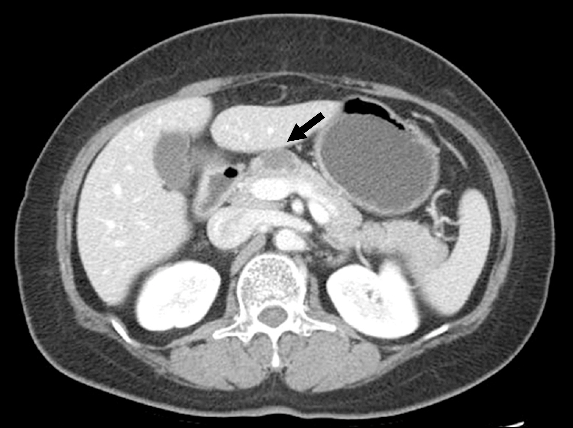
Fig. 1. Abdominal CT finding revealed a 2.2×1.2 cm cystic mass in the pancreas body (arrow).

Fig. 2. Follow-up abdominal CT finding 9 months after initial CT. It revealed a 3.3×2.2 cm cystic mass in the same area (arrow).
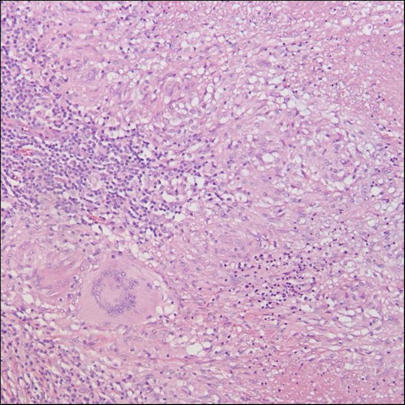
Fig. 3. The microscopic finding of resected specimen revealed chronic granulomatous inflammation with caseous necrosis and multinucleated giant cells (H&E stain, ×200).
Reference
-
1. Bhansali S. Abdominal tuberculosis. Experience with 300 cases. Am J Gastroenterol. 1977; 67:324–327.2. Auerbach O. Acute generalized miliary tuberculosis. Am J Pathol. 1944; 20:121–136.3. Hulnick DH, Megibow AJ, Naidich DP, Hilton S, Cho KC, Balthazar EJ. Abdominal tuberculosis: CT evaluation. Radiology. 1985; 157:199–204.
Article4. Yokoyama T, Miyagawa S, Noike T, Shimada R, Kawasaki S. Isolated pancreatic tuberculosis. Hepatogastroenterology. 1999; 46:2011–2014.5. Woodfield JC, Windsor JA, Godfrey CC, Orr DA, Officer NM. Diagnosis and management of isolated pancreatic tuberculosis: recent experience and literature review. ANZ J Surg. 2004; 74:368–371.
Article6. Ko YW, Hwang EH, Lee DW. Obstructive jaundice due to tuberculous lymphadenitis and/or tuberculosis of the pancreas: a case report. Korean J Gastroenterol. 1976; 8:67–75.7. Choi HH, Song TJ, Paik SH, et al. A case of pancreatic tuberculosis. Korean J Gastroenterol. 1988; 20:740–745.8. Park JM, Song SY, Park SW, et al. A case of intraabdominal tuberculosis with pancreatic involvement showing interesting ERP finding. Korean J Gastrointest Endosc. 1995; 15:285–293.9. Lee S, Choi SH, Yun SS, Lim KW. Tuberculous pancreatic abscess: a case report. Korean J Hepatobiliary Pancreat Surg. 1999; 3:211–214.10. Park JW, Kim DI, Park SS, et al. Two cases of pancreatic tuberculosis masquerading as a pancreatic mass. Korean J Gastroenterol. 2000; 35:671–675.11. Park HJ, Nam SW, Roe IW, et al. A case of pancreatic tuberculosis diagnosed by fine needle aspiration. Korean J Gastrointest Endosc. 2001; 38:57–60.12. Yoo DK, Cho JS, Shin KS, Kang DY. Tuberculous abscess of the pancreas presenting as obstructive jaundice: a case report. J Korean Radiol Soc. 2002; 46:593–595.
Article13. Lee HY, Kim SH, Park KJ, Kim YH, Lee JH, Roh MS. Pancreatic tuberculosis with microcystic adenoma. J Korean Surg Soc. 2005; 68:522–525.14. Hwang LS, Nam SW, Lee SE, et al. A case of pancreaticobiliary duct obstruction due to pancreatic tuberculosis combined with a colon adenocarcinoma and tuberculous colitis. Korean J Gastrointest Endosc. 2007; 35:267–271.15. Knowles KF, Saltman D, Robson HG, Lalonde R. Tuberculous pancreatitis. Tubercle. 1990; 71:65–68.
Article16. Stock KP, Riemann JF, Stadler W, Rösch W. Tuberculosis of the pancreas. Endoscopy. 1981; 13:178–180.
Article17. Warshaw AL, Brugge WR, Lewandrowski KB, Pitman MB. Case records of the Massachusetts General Hospital. Weekly clinicopathological exercises. Case 35-2003. A 75-year-old man with a cystic lesion of the pancreas. N Engl J Med. 2003; 349:1954–1961.18. Bhatia V, Garg PK, Arora VK, Sharma R. Isolated pancreatic tuberculosis mimicking intraductal pancreatic mucinous tumor. Gastrointest Endosc. 2008; 68:610–611.
Article19. Nakai Y, Tsujino T, Kawabe T, et al. Pancreatic tuberculosis with a pancreaticobiliary fistula. Dig Dis Sci. 2007; 52:1225–1228.
Article20. Liu Q, He Z, Bie P. Solitary pancreatic tuberculous abscess mimicking pancreatic cystadenocarcinoma: a case report. BMC Gastroenterol. 2003; 3:1.
Article21. Fischer G, Spengler U, Neubrand M, Sauerbruch T. Isolated tuberculosis of the pancreas masquerading as a pancreatic mass. Am J Gastroenterol. 1995; 90:2227–2230.22. Rezeig MA, Fashir BM, Al-Suhaibani H, Al-Fadda M, Amin T, Eisa H. Pancreatic tuberculosis mimicking pancreatic carcinoma: four case reports and review of the literature. Dig Dis Sci. 1998; 43:329–331.23. Takhtani D, Gupta S, Suman K, et al. Radiology of pancreatic tuberculosis: a report of three cases. Am J Gastroenterol. 1996; 91:1832–1834.24. Ahlawat SK, Charabaty-Pishvaian A, Lewis JH, Haddad NG. Pancreatic tuberculosis diagnosed with endoscopic ultrasound guided fine needle aspiration. JOP. 2005; 6:598–602.25. Kaushik N, Schoedel K, McGrath K. Isolated pancreatic tuberculosis diagnosed by endoscopic ultrasound-guided fine needle aspiration: a case report. JOP. 2006; 7:205–210.26. Itaba S, Yoshinaga S, Nakamura K, et al. Endoscopic ultrasound-guided fine-needle aspiration for the diagnosis of peri-pancreatic tuberculous lymphadenitis. J Gastroenterol. 2007; 42:83–86.
Article
- Full Text Links
-
- Actions
-
Cited
- CITED
-
- Close
- Share
-
- Similar articles
-
- A Case of Mucinous Cystic Adenocarcinoma of the Pancreas
- Pancreatic Schwannoma with Cystic Degeneration: A Case Report and Literature Review
- Tuberculous Abscess of the Pancreas Presenting as Obstructive Jaundice: A Case Report
- Pancreatic Collision Tumor of Desmoid-Type Fibromatosis and Mucinous Cystic Neoplasm: A Case Report
- Pathologic Features of Pancreatic Cystic Neoplasms
