Korean J Radiol.
2002 Mar;3(1):45-48. 10.3348/kjr.2002.3.1.45.
The Usefulness of Pulsatile Flow Detection in Measuring Resistive Index in Renal Doppler US
- Affiliations
-
- 1Department of Radiology, Seoul National University College of Medicine and the Institute of Radiation Medicine, SNUMRC, Seoul National University Hospital, Seoul, Korea. pinksu77@korea.com
- KMID: 1758446
- DOI: http://doi.org/10.3348/kjr.2002.3.1.45
Abstract
OBJECTIVE
To assess the usefulness of pulsatile flow detection (PFD), a newly developed function of color Doppler US, in measuring resistive index (RI) in renal Doppler US and to compare it with conventional color Doppler (CCD).
MATERIALS AND METHODS
Fifty-six kidneys in 31 patients were randomly selected and divided into two groups. In group A, RI was measured first with the aid of CCD, and then with PFD. In group B, data were obtained in the reverse order. The time required for each RI measurement was recorded in seconds. The quality of the Doppler spectral waveform was subjectively graded as 0, 1, or 2 and examination time and waveform quality were compared between PFD and CCD.
RESULTS
The time required to measure RI with PFD (PFD time) was less than with CCD (CCD time) (mean 42.7 secs vs. mean 70.3 secs; p = 0.031). There was no significant difference in PFD time between group A and B, but CCD time was shorter in group B (70.3 secs vs. 24.6 secs; p = 0.0004). Spectral waveform quality was not significantly different between PFD and CCD.
CONCLUSION
The time required to measure RI in kidneys can be shortened with the aid of the PFD function in color Doppler US without affecting the quality of the examination.
MeSH Terms
Figure
-

Fig. 1 A. PFD image of a kidney. Interlobar arteries are colored green (arrows) and easily distinguished from other non-pulsatile flows. B. The Doppler cursor is located on a green-colored interlobar artery and clear spectral waveforms (grade 2) are obtained.
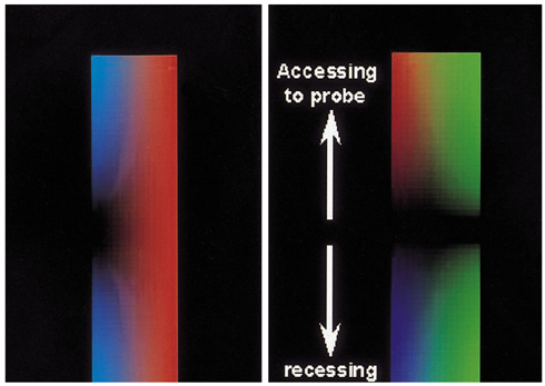
Fig. 2 PFD color modes. In the two-color mode (left), all pulsatile flows are red, while nonpulsatile flows are blue. In the three-color mode (right), pulsatile flows are green, and are added to colors depicted by directional color Doppler (red / blue).
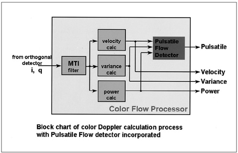
Fig. 3 Principle of PFD. A 'pulsatile flow detector' is placed in a color flow processor and flow pulsatility is determined as a function of velocity difference, variance, and power.
Reference
-
1. Platt JF. Doppler ultrasound of the kidney. Semin Ultrasound CT MR. 1997. 18:22–32.2. Lee HJ, Kim SH, Jeong YK, Yeon KM. Doppler sonographic resistive index in obstructed kidneys. J Ultrasound Med. 1996. 15:613–618.3. Tublin ME, Dodd GD III. Sonography of renal transplantation. Radiol Clin North Am. 1995. 33:447–459.4. Keogan MT, Kliewer MA, Hertzberg BS, DeLong DM, Tupler RH, Carroll BA. Renal resistive indexes: variability in Doppler US measurement in a healthy population. Radiology. 1996. 199:165–169.5. Eibenberger K, Schima H, Trubel W, Scherer R, Dock W, Grabenwoger F. Intrarenal Doppler ultrasonography: which vessel should be investigated? J Ultrasound Med. 1995. 14:451–455.6. Seong CK, Kim SH, Sim JS. Detection of segmental branch renal artery stenosis by Doppler US: A Case Report. Korean Journal of Radiology. 2001. 2:57–60.7. Mori H, Haneki H, Yamakawa T, et al. Pulsatile flow detection for evaluation of tumor vessels of hepatocellular carcinoma. Nippon Shokakibyo Gakkai Zasshi. 2000. 97:484.
- Full Text Links
-
- Actions
-
Cited
- CITED
-
- Close
- Share
-
- Similar articles
-
- Evaluation of Factors Affecting the Renal Doppler Waveform with the Use of an Electrical Circuit Model
- Pelvis dilatation and mucosal thickening of transplanted kidney: comparative study of resistive index and ultrasonographic finding
- Resistive Index Analysis Using Doppler Ultrasonography in Patients with Acute Stroke
- Usefulness of resistive index on spectral Doppler ultrasonography in the detection of renal cell carcinoma in patients with end-stage renal disease
- Correlation between the Resistive Index of the Doppler Sonography and Serum Creatinine Level in Experimentally Induced Crescentic Glomerulonephritis in Rabbits
