J Korean Soc Spine Surg.
2008 Sep;15(3):190-193. 10.4184/jkss.2008.15.3.190.
Paraparesis due to Posterior Migration of Ruptured Disc in the Adjacent Segment after Spinal Fusion: Unusual Junctional Problem
- Affiliations
-
- 1Department of Orthopaedic Surgery, Hanyang University Guri Hospital, Guri, Korea. hyparkys@hanyang.ac.kr
- 2Department of Pathology, Hanyang University Guri Hospital, Guri, Korea.
- 3Department of Orthopaedic Surgery, College of Medicine, Hanyang University, Seoul, Korea.
- KMID: 1747203
- DOI: http://doi.org/10.4184/jkss.2008.15.3.190
Abstract
- Posterior epidural migration of sequestered lumbar disc fragments is an uncommon event. We present here an especially uncommon case involving a patient with paraparesis that was due to posterior migration of a ruptured disc in the adjacent segment after spinal fusion. The patient had a herniated lumbar disc in a diseased spinal junction with sequestered fragments that were located posterior to the thecal sac.
Keyword
MeSH Terms
Figure
-
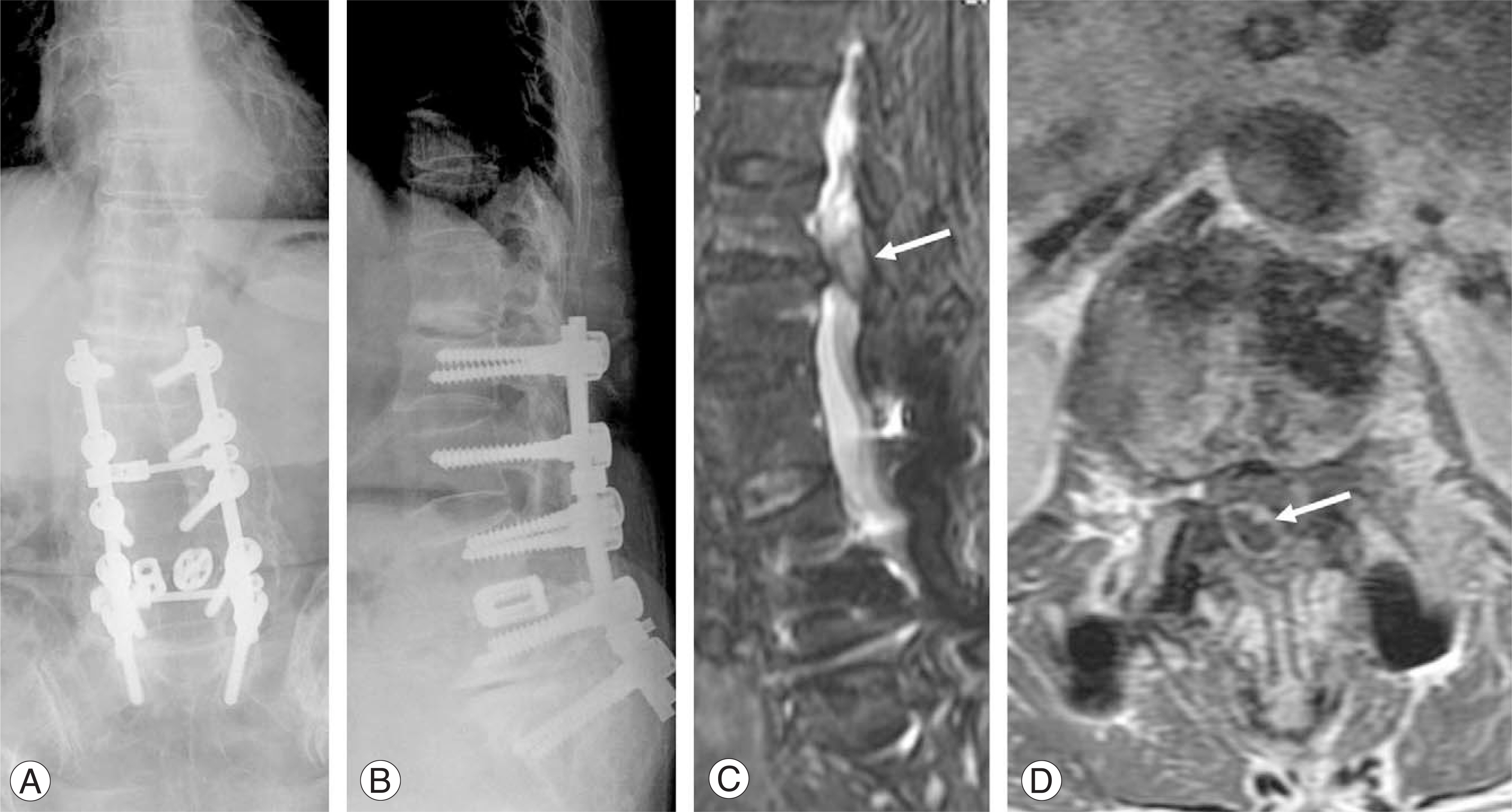
Fig. 1. (A) Initial x-ray of the thoracolumbar spine shows junctional problem at L1-L2 level. (B) Preoperative thoracolumbar spine lateral view shows junctional problem. (C) Posterior epidural disc fragment is detected with mild enhancement on sagittal MR enhancement image (arrow) (D) Margin of posterior epidural disc fragment is seen with enhancement at the posterior to thecal sac of L1-2 intervertebral disc space on axial MR enhancement image (arrow)
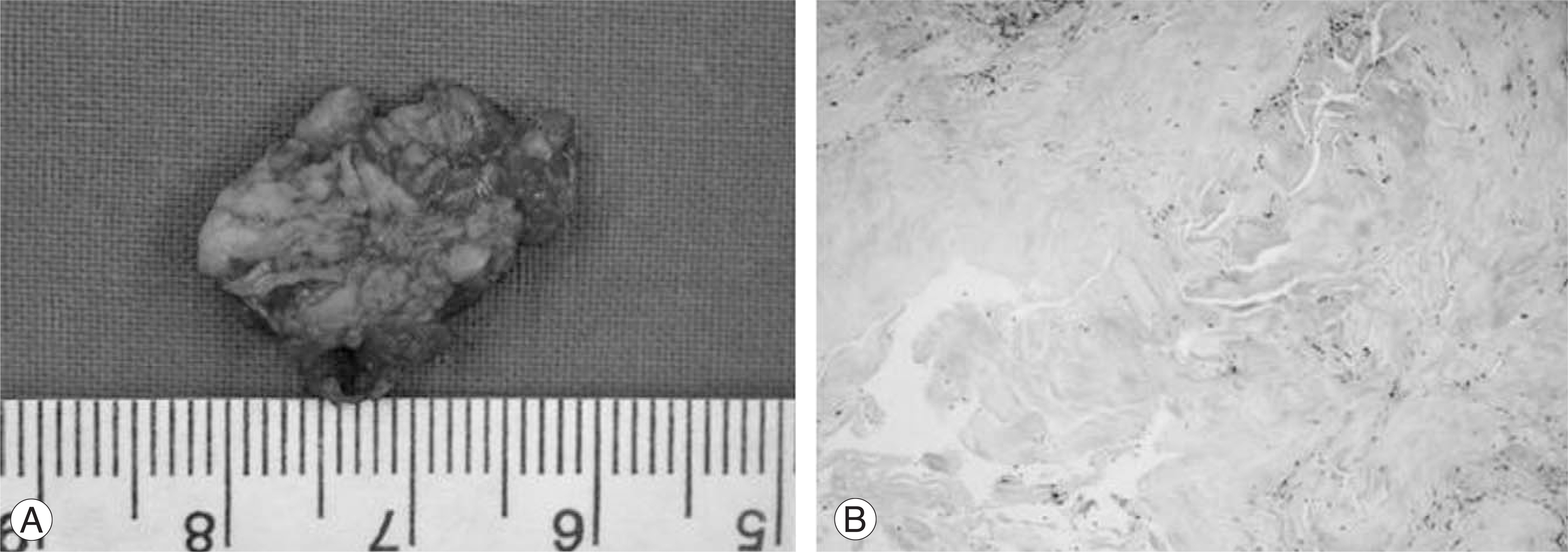
Fig. 2. (A) Intraoperative photography shows 1.5×1.8×2.0 cm-sized posteriorly migrated extradural disc fragment. (B) Photomicrograph shows disc degeneration (HE stain ×100)
Reference
-
1). Bonaroti EA, Welch WC. Posterior epidural migration of an extruded lumbar disc fragment causing cauda equina syndrome. Clinical and magnetic resonance imaging evaluation. Spine. 1998; 23:378–381.2). Manabe S, Tateishi A. Epidural migration of extruded cervical disc and its surgical treatment. Spine. 1986; 11:873–878.
Article3). Giannini C, Scheithauer BW, Wenger DE, Unni KK. Pigmented villonodular synovitis of the spine: a clinical, radiological and morphological study of 12 cases. J Neurosurg. 1996; 84:592–597.
Article4). Baker JK, Hanson GW. Cyst of the ligamentum flavum. Spine. 1994; 19:1092–1094.
Article5). Masaryk TJ, Ross JS, Modic MT, Boumphrey F, Bohlman H, Wilber G. High-resolution MR imaging of sequestered lumbar intervertebral disks. Am I Roentgenol. 1988; 150:1155–1162.
Article6). Schellinger D, Manz HJ, Vidic B, et al. Disk fragment migration. Radiology. 1990; 175:831–836.
Article7). Lutz JD, Smith RR, Jones HM. CT myelography of a fragment of a lumbar disk sequestered posterior to the thecal sac. AJNR Am J Neuroradiol. 1990; 11:610–611.8). Bonaldi VM, Duong H, Starr MR, Sarazin L, Richardson J. Tophaceous gout of the lumbar spine mimicking an epidural abscess: MR features. AJNR Am J Neuroradiol. 1996; 17:1949–1952.9). Liu SS, Williams KD, Drayer BP, Spetzler RF, Sonntag VK. Synovial cysts of the lumbosacral spine: diagnosis by MR imaging. AJNR Am J Neuroradiol. 1989; 10:1239–1242.
Article
- Full Text Links
-
- Actions
-
Cited
- CITED
-
- Close
- Share
-
- Similar articles
-
- Acute Cervical Myelopathy Due to Ruptured Disc During Leisure Sports Activity in Adjacent Segment
- Surgical Treatment of Adjacent Segment Degeneration after Spinal Fusion in Degenerative Lumbar Disc Disease
- Acute Paraplegia Secondary to Thoracic Disc Herniation of the Adjacent Segment Following Thoracolumbar Fusion and Instrumentation
- Revision Surgery for Spinal Stenosis Developed at the Adjacent Segment after Lumbar Fusion
- A Comparison of Adjacent Segment Diseases Above One Versus Above Two Vertebral Segment after Spinal Fusion of the Degenerative Lumbar Disease
