Evaluation of Deep Vein Thrombosis with Multidetector Row CT after Orthopedic Arthroplasty: a Prospective Study for Comparison with Doppler Sonography
- Affiliations
-
- 1Department of Radiology, Inha University, College of Medicine, Incheon, Korea.
- 2Department of Radiology, Gachon University, Gil Medical Center, Incheon, Korea. ho7ok7@gilhospital.com
- KMID: 1734276
- DOI: http://doi.org/10.3348/kjr.2008.9.1.59
Abstract
OBJECTIVE
This prospective study evaluated the ability of indirect 16-row multidetector CT venography, in comparison with Doppler sonography, to detect deep vein thrombosis after total hip or knee replacement. MATERIALS AND METHODS: Sixty-two patients had undergone orthopedic replacement surgery on a total of 30 hip joints and 54 knee joints. The CT venography (scan delay time: 180 seconds; slice thickness/increment: 2/1.5 mm) and Doppler sonography were performed 8 to 40 days after surgery. We measured the z-axis length of the beam hardening artifact that degraded the image quality so that the presence of deep vein thrombosis couldn't be evaluated on the axial CT images. The incidence and location of deep vein thrombosis was analyzed. The diagnostic performance of the CT venograms was evaluated and compared with that of Doppler sonography as a standard of reference. RESULTS: The z-axis length (mean +/- standard deviation) of the beam hardening artifact was 4.5 +/- 0.8 cm in the arthroplastic knees and 3.9 +/- 2.9 cm in the arthroplastic hips. Deep vein thrombosis (DVT) was found in the popliteal or calf veins on Doppler sonography in 30 (48%) of the 62 patients. The CT venography has a sensitivity, specificity, positive predictive value, negative predictive value and accuracy of 90%, 97%, 96%, 91% and 94%, respectively. CONCLUSION: The ability of CT venography to detect DVT was comparable to that of Doppler sonography despite of beam hardening artifact. Therefore, CT venography is feasible to use as an alternative modality for evaluating post-arthroplasty patients.
MeSH Terms
-
Adult
Aged
Aged, 80 and over
*Arthroplasty, Replacement, Hip
*Arthroplasty, Replacement, Knee
Artifacts
Female
Humans
Leg/*blood supply
Male
Middle Aged
Predictive Value of Tests
Prospective Studies
Sensitivity and Specificity
Tomography, X-Ray Computed/*methods
Venous Thrombosis/etiology/*radiography/*ultrasonography
Figure
-

Fig. 1 A 63-year-old female underwent left total knee arthroplasty. A. Note the degradation of the image quality of the 3.5 cm-long segment (between the two arrows) in the popliteal fossa on the three-dimensional volume rendered image. B. An axial CT image shows the non-enhancing, low-attenuated lesions surrounded by contrast material within the left calf veins (asterisks). C. Color Doppler sonography reveals a hypoechoic lesion partially obstructing the left calf vein. There were blood flow signals surrounding the lesion.

Fig. 2 A 46-year-old female underwent left total hip arthroplasty. A. The three-dimensional volume rendering image shows the degradation of image quality along the long segment (between the two arrows) due to beam hardening artifact by the arthroplastic joint material. However, the entire common and superficial femoral veins, except for a 2 cm-long segment, could be evaluated for whether deep vein thrombosis was present or not. B. An axial CT image shows beam hardening artifact traversing the adjacent superficial femoral vein (arrow). C. A coronal image shows non-enhancing, low-attenuated lesions surrounded by contrast material within the left calf veins (arrows). Doppler sonography confirmed the deep vein thrombosis (not shown).

Fig. 3 A 54-year-old female underwent left total hip and knee arthroplasty simultaneously. A, B. Doppler sonography without (A) and with compression (B) revealed deep vein thrombosis in the left calf vein. The blood flow signal means partial occlusion of the left calf veins. After compression, the deep vein thrombosis lesion showed no compressibility. However, deep vein thrombosis was initially undetected on the CT venograms. C. On the retrospective analysis, a coronal CT image showed a small, non-occlusive thrombus (arrows) within the left calf vein.
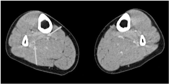
Fig. 4 A 32-year-old female underwent right total hip arthroplasty. An axial CT image showed non-enhancing, low-attenuated lesions surrounded by high-attenuated tissue in the right calf (arrows). However, there was no definite evidence of deep vein thrombosis in the right calf vein despite the repeated sonographic examinations. This was a false-positive case.
Cited by 3 articles
-
Venous Thromboembolism in Korean Patients Undergoing Major Orthopedic Surgery: A Prospective Observational Study using Computed Tomographic (CT) Pulmonary Angiography and Indirect CT Venography
Seung-Ick Cha, Shin-Yeop Lee, Chang-Ho Kim, Jae-Yong Park, Tae-Hoon Jung, Jae-Hyuck Yi, Jongmin Lee, Seung Huh, Hyun-Joo Lee, Shin-Yoon Kim
J Korean Med Sci. 2010;25(1):28-34. doi: 10.3346/jkms.2010.25.1.28.Comparison of Image Qualities of 80 kVp and 120 kVp CT Venography Using Model-Based Iterative Reconstruction at Same Radiation Dose
Hyun Jung Baek, Ki Seok Choo, Kyung Jin Nam, Jae-Yeon Hwang, Ji Won Lee, Jin You Kim, Hyuk Jae Jung
J Korean Soc Radiol. 2018;78(4):235-241. doi: 10.3348/jksr.2018.78.4.235.Usefulness of the Computed Tomography Venography for Evaluation of Leg Edema Including Deep Vein Thrombosis in Rehabilitation Patients
Ji Hea Chang, Ho Jun Lee, Jae Hyun Kwon, Gi Hyeong Ryu, Heebong Moon, Changjae Kim, Ki Yeon Nam, Bum Sun Kwon
Ann Rehabil Med. 2014;38(6):812-820. doi: 10.5535/arm.2014.38.6.812.
Reference
-
1. Elias A, Cadene A, Elias M, Puget J, Tricoire JL, Colin C, et al. Extended lower limb venous ultrasound for the diagnosis of proximal and distal vein thrombosis in asymptomatic patients after total hip replacement. Eur J Vasc Endovasc Surg. 2004. 27:438–444.2. Nathan S, Aleem MA, Thiagarajan P, Das De S. The incidence of proximal deep vein thrombosis following total knee arthroplasty in an Asian population: a Doppler ultrasound study. J Orthop Surg (Hong Kong). 2003. 11:184–189.3. Della Valle CJ, Steiger DJ, DiCesare PE. Duplex ultrasonography in patients suspected of postoperative pulmonary embolism following total joint arthroplasty. Am J Orthop. 2003. 32:386–388.4. Sudo A, Sano T, Horikawa K, Yamakawa T, Shi D, Uchida A. The incidence of deep vein thrombosis after hip and knee arthroplasties in Japanese patients: a prospective study. J Orthop Surg (Hong Kong). 2003. 11:174–177.5. Cordell-Smith JA, Williams SC, Harper WM, Gregg PJ. Lower limb arthroplasty complicated by deep venous thrombosis. Prevalence and subjective outcome. J Bone Joint Surg Br. 2004. 86:99–101.6. Kim YH, Kim JS. Incidence and natural history of deep-vein thrombosis after total knee arthroplasty. J Bone Joint Surg Br. 2002. 84:566–570.7. Kim YH, Oh SH, Kim JS. Incidence and natural history of deepvein thrombosis after total hip arthroplasty. A prospective and randomised clinical study. J Bone Joint Surg Br. 2003. 85:661–665.8. Colwell CW Jr. Managing thromboembolic risk in hip and knee arthroplasty: state of the art. Orthopedics. 2003. 26:S231–S236.9. Lieberman JR, Hsu WK. Prevention of venous thromboembolic disease after total hip and knee arthroplasty. J Bone Joint Surg Am. 2005. 87:2097–2112.10. Mehta JS, Nicolaou N, Kiryluk S, Fordyce MJ. Venous leg ulcers after hip replacement. A clinical evaluation at 5 to 12 years. J Bone Joint Surg Br. 2003. 85:960–962.11. Lee AY, Gent M, Julian JA, Bauer KA, Eriksson BI, Lassen MR, et al. Bilateral vs. ipsilateral venography as the primary efficacy outcome measure in thromboprophylaxis clinical trials: a systematic review. J Thromb Haemost. 2004. 2:1752–1759.12. Wang CJ, Wang JW, Weng LH, Hsu CC, Lo CF. Outcome of calf deep-vein thrombosis after total knee arthroplasty. J Bone Joint Surg Br. 2003. 85:841–844.13. Walker RH. Secondary prevention of venous thromboembolism in joint replacement using duplex ultrasonography. Orthopedics. 1994. 17:14–17.14. Leutz DW, Stauffer ES. Color duplex Doppler ultrasound scanning for detection of deep venous thrombosis in total knee and hip arthroplasty patients. Incidence, location, and diagnostic accuracy compared with ascending venography. J Arthroplasty. 1994. 9:543–548.15. Davidson BL, Elliott CG, Lensing AW. Low accuracy of color Doppler ultrasound in the detection of proximal leg vein thrombosis in asymptomatic high-risk patients. The RD Heparin Arthroplasty Group. Ann Intern Med. 1992. 117:735–738.16. Elliott CG, Suchyta M, Rose SC, Talbot S, Ford C, Raskob G, et al. Duplex ultrasonography for the detection of deep vein thrombi after total hip or knee arthroplasty. Angiology. 1993. 44:26–33.17. Garino JP, Lotke PA, Kitziger KJ, Steinberg ME. Deep venous thrombosis after total joint arthroplasty. The role of compression ultrasonography and the importance of the experience of the technician. J Bone Joint Surg Am. 1996. 78:1359–1365.18. Ciccone WJ 2nd, Fox PS, Neumyer M, Rubens D, Parrish WM, Pellegrini VD Jr. Ultrasound surveillance for asymptomatic deep venous thrombosis after total joint replacement. J Bone Joint Surg Am. 1998. 80:1167–1174.19. Eskandari MK, Sugimoto H, Richardson T, Webster MW, Makaroun MS. Is color-flow duplex a good diagnostic test for detection of isolated calf vein thrombosis in high-risk patients. Angiology. 2000. 51:705–710.20. Robinson KS, Anderson DR, Gross M, Petrie D, Leighton R, Stanish W, et al. Ultrasonographic screening before hospital discharge for deep venous thrombosis after arthroplasty: the post-arthroplasty screening study. Ann Intern Med. 1997. 127:439–445.21. Grady-Benson JC, Oishi CS, Hanson PB, Colwell CW Jr, Otis SM, Walker RH. Routine postoperative duplex ultrasonography screening and monitoring for the detection of deep vein thrombosis. A survey of 110 total hip arthroplasties. Clin Orthop Relat Res. 1994. 307:130–141.22. Kalodiki E, Nicolaides AN, Al-Kutoubi A, Cunningham DA, Crofton M. Duplex scanning in the postoperative surveillance of patients undergoing total hip arthroplasty. J Arthroplasty. 1997. 12:310–316.23. Robinson KS, Anderson DR, Gross M, Petrie D, Leighton R, Stanish W, et al. Accuracy of screening compression ultrasonography and clinical examination for the diagnosis of deep vein thrombosis after total hip or knee arthroplasty. Can J Surg. 1998. 41:368–373.24. Westrich GH, Allen ML, Tarantino SJ, Ghelman B, Schneider R, Laskin RS, et al. Ultrasound screening for deep venous thrombosis after total knee arthroplasty: 2-year reassessment. Clin Orthop Relat Res. 1998. 356:125–133.25. Verlato F, Bruchi O, Prandoni P, Camporese G, Maso G, Busonera F, et al. The value of ultrasnoud screening for proximal vein thrombosis after total hip arthroplasty. Thromb Haemost. 2001. 86:534–537.26. Schwarcz TH, Matthews MR, Hartford JM, Quick RC, Kwolek CJ, Minion DJ, et al. Surveillance venous duplex is not clinically useful after total joint arthroplasty when effective deep venous thrombosis prophylaxis is used. Ann Vasc Surg. 2004. 18:193–198.27. Westrich GH, Salvati EA, Sharrock N, Potter HG, Sanchez PM, Sculco TP. The effect of intraoperative heparin administered during total hip arthroplasty on the incidence of proximal deep vein thrombosis assessed by magnetic resonance venography. J Arthroplasty. 2005. 20:42–50.28. Pookarnjanamorakot C, Sirisriro R, Eurvilaichit C, Jaovisidha S, Koysombatolan I. The incidence of deep vein thrombosis and pulmonary embolism after total knee arthroplasty: the screening study by radionuclide venography. J Med Assoc Thai. 2004. 87:869–876.29. Szapiro D, Ghaye B, Willems V, Zhang L, Albert A, Dondelinger RF. Evaluation of CT time-density curves of lowerlimb veins. Invest Radiol. 2001. 36:164–169.30. Garg K, Kemp JL, Wojcik D, Hoehn S, Johnston RJ, Macey LC, et al. Thromboembolic disease: comparison of combined CT pulmonary angiography and venography with bilateral leg sonography in 70 patients. AJR Am J Roentgenol. 2000. 175:997–1001.31. Duwe KM, Shiau M, Budorick NE, Austin JH, Berkmen YM. Evaluation of the lower extremity veins in patients with suspected pulmonary embolism: a retrospective comparison of helical CT venography and sonography. AJR Am J Roentgenol. 2000. 175:1525–1531.32. Begemann PGC, Bonacker M, Kemper J, Guthoff AE, Hahn KE, Steiner P, et al. Evaluation of the deep venous system in patients with suspected pulmonary embolism with multidetector CT: a prospective study in comparison to Doppler sonography. J Comput Assist Tomogr. 2003. 27:399–409.33. Ghaye B, Szapiro D, Willems V, Dondelinger RF. Pitfalls in CT venography of lower limbs and abdominal veins. AJR Am J Roentgenol. 2002. 178:1465–1471.34. Delis KT, Hunt N, Strachan RK, Nicolaides AN. Incidence, natural history and risk factors of deep vein thrombosis in elective knee arthroscopy. Thromb Haemost. 2001. 86:817–821.
- Full Text Links
-
- Actions
-
Cited
- CITED
-
- Close
- Share
-
- Similar articles
-
- Evaluation of Deep Vein Thrombosis with Multidetector Row CT after Orthopedic Arthroplasty: a Prospective Study for Comparison with Doppler Sonography
- The Diagnostic Availability of Multidetector-Row Computed Tomography(MDCT) in Deep Vein Thrombosis Developed after Joint Arthroplasty
- The Prevalence and Surveillance of Deep Vein Thrombosis after Total Hip Arthroplasty
- Diagnosis of the Deep Vein Thrombosis with Multidetector-Row Computed Tomographic Venography after Total Knee Arthroplasty
- The Effects of Foot Pump on Prevention of Deep Vein Thrombosis Following Total Knee Arthroplasty
