Yonsei Med J.
2005 Apr;46(2):296-299. 10.3349/ymj.2005.46.2.296.
Bilateral Congenital Anophthalmos and Agenesis of the Optic Pathways
- Affiliations
-
- 1Department of Anatomy, Mersin University, Faculty of Medicine, Mersin, Turkey. maktekin@mersin.edu.tr
- 2Department of Ophthalmology, Mersin University, Faculty of Medicine, Mersin, Turkey.
- 3Radiodiagnostic University IMC Hospital, Turkey.
- KMID: 1734059
- DOI: http://doi.org/10.3349/ymj.2005.46.2.296
Abstract
- This report presents a rare example of a bilateral congenital anophthalmos and an agenesis of the optic pathways. The MR imaging studies revealed that the eyeballs, optic nerves, optic chiasm, optic tracts and optic radiation were absent. The chromosomal examination was normal. Mild mental retardation was also observed. Apart from the rarity of the anophthalmos and the total absence of the optic pathways, no etiologic reason for this pathology could be detected, which makes this case more significant.
Keyword
MeSH Terms
Figure
-
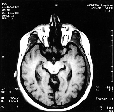
Fig. 1 Axial T1-weighted MR image shows bilateral fibrotic eye bulbs in both orbits (arrow). The rudimental medial and lateral recti are medially displaced (arrow head). Minimal enlargement of the lateral ventricles but no hypotense signal changes of the optic radiation can be observed.

Fig. 2 Inversion recovery sequence shows the enlarged ventricles and an absence of the optic radiations (arrow).

Fig. 3 Midsagittal T1-weighted MR image shows an absence of the optic chiasm (arrow) and a normal corpus callosum.
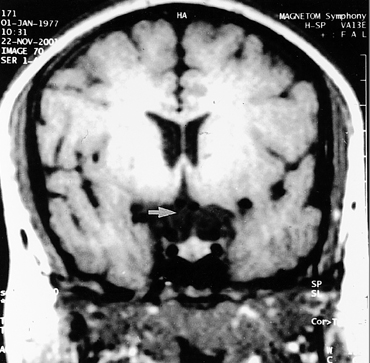
Fig. 4 Coronal T1-weighted MR image shows an absence of the optic chiasm (arrow).
Reference
-
1. BenEzra D, Sela M, Peer J. Bilateral anophthalmia and unilateral microphthalmia in two siblings. Ophthalmologica. 1989. 198:140–144.2. Albernaz SA, Castillo M, Hudgins PA, Mukherji SK. Imaging findings in patients with clinical anophthalmos. AJNR Am J Neuroradiol. 1997. 18:112–113.3. Brunquell PJ, Papale JH, Horton JC, Williams RS, Zgrabik MJ, Albert DM, et al. Sex-linked hereditary bilateral anophthalmos. Arch Ophthalmol. 1984. 102:108–113.4. Marcus DM, Shore JW, Albert DM. Anophthalmia in the focal dermal hypoplasia syndrome. Arch Ophthalmol. 1990. 108:96–100.5. Sener RN. Cranial MR imaging findings in Waardenburg syndrome: anophthalmia, and hypothalamic hamartoma. Comput Med Imaging Graph. 1998. 22:409–411.6. Moore KL, Persaud TVN. The developing human. 1998. 6th ed. Philadelphia: W.B. Saunders Company.7. Sensi A, Incorvaia C, Sebastiani A, Calzolari E. Clinical anophthalmos in a family. Clin Genet. 1987. 32:156–159.8. Daxecker F, Felber S. Magnetic resonance imaging features of congenital anophthalmia. Ophthalmologica. 1993. 206:139–142.9. Scott IU, Warman R, Altman N. Bilateral aplasia of the optic nerves, chiasm, and tracts in an otherwise healthy infant. Am J Ophthalmol. 1997. 124:409–410.10. Waheed K, Jan W, Calver DM. Aplasia of the optic chiasm and tracts with unifocal polymicrogyria in an otherwise healthy infant. J Pediatr Ophthalmol Strabismus. 2002. 39:187–189.11. Pearce WG, Nigam S, Rootman J. Primary anophthalmos. Histological and genetic features. Can J Ophthalmol. 1974. 9:141–145.
