Advanced Cardiac MR Imaging for Myocardial Characterization and Quantification: T1 Mapping
- Affiliations
-
- 1Department of Radiology, Yonsei University College of Medicine, Seoul, Korea. bchoi@yuhs.ac
- KMID: 1722994
- DOI: http://doi.org/10.4070/kcj.2013.43.1.1
Abstract
- Magnetic resonance as an imaging modality provides an excellent soft tissue differentiation, which is an ideal choice for cardiac imaging. Cardiac magnetic resonance (CMR) allows myocardial tissue characterization, as well as comprehensive evaluation of the structures. Although late gadolinium enhancement after injection of the gadolinium extracellular contrast agent has further extended our ability to characterize the myocardial tissue, it also has limitations in the quantification of enhanced myocardial tissue pathology, and the detection of diffuse myocardial disease, which is not easily recognized by enhancement contrast. Recently, the remarkable advances in CMR technique, such as T1 mapping, which can quantitatively evaluate myocardial status, showed potentials to overcome limitations of existing CMR sequences and to expand the application of CMR. This article will review the technical and clinical points to be considered in the practical use of pre- and post-contrast T1 mapping.
MeSH Terms
Figure
-
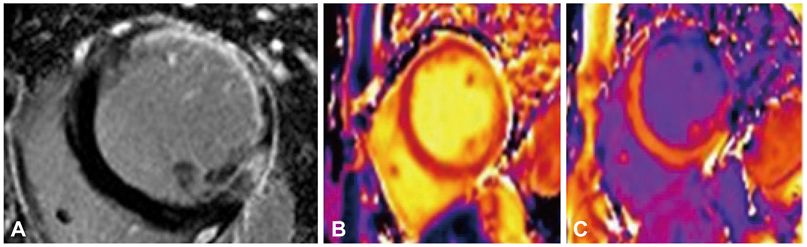
Fig. 1 Late gadolinium enhancement (LGE) (A), pre- and post-contrast (B and C) T1 map images using 3-T MR system in a patients with prior myocardial infarction in the lateral wall. LGE-MR image shows strong subendocardial enhancement and diffuse wall thinning from chronic infarction in the left anterior descending artery territory of the middle left ventricle level. Post-contrast T1 map image shows a marked T1 shortening in the peri-infarct region, which is not grossly enhanced on LGE image, when compared to the remote zone.
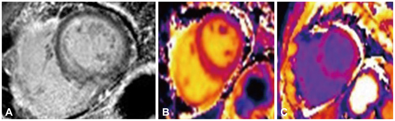
Fig. 2 Late gadolinium enhancement (LGE) (A), pre- and post-contrast (B and C) T1 map images using 3-T MR system in a patient with secondary myocardial amyloidosis from multiple myeloma. LGE-MR image shows diffuse subendocardial gadolinium enhancement, which is a typical finding of myocardial amyloidosis deposition. Post-contrast T1 map image shows decrease of T1 value in the myocardium which enables quantification of extracellular volume fraction.
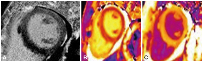
Fig. 3 Late gadolinium enhancement (LGE) (A), pre- and post-contrast (B and C) T1 map images using 3-T MR system in a patient with esoniophilic myocarditis diagnosed by the biopsy. LGE-MR image shows no remarkable late gadolinium enhancement in the myocardium. Pre-contrast T1 map image shows the prolongation of T1 value in the focal area of mid ventricular septum, compared to the lateral wall. It can suggest the probable edema in the myocardium. Post-contrast T1 mapping show diffusely low T1 value (mean±standard deviation, 410.4±34.4 msec) from gadolinium-induced T1 shortening in the myocardium. Myocardial extracellular volume fraction calculated by the T1 mapping results is 33.5% which is higher than that in normal control in the literature.
Reference
-
1. Bottomley PA, Foster TH, Argersinger RE, Pfeifer LM. A review of normal tissue hydrogen NMR relaxation times and relaxation mechanisms from 1-100 MHz: dependence on tissue type, NMR frequency, temperature, species, excision, and age. Med Phys. 1984. 11:425–448.2. Cummings KW, Bhalla S, Javidan-Nejad C, Bierhals AJ, Gutierrez FR, Woodard PK. A pattern-based approach to assessment of delayed enhancement in nonischemic cardiomyopathy at MR imaging. Radiographics. 2009. 29:89–103.3. Callot V, Galanaud D, Figarella-Branger D, et al. Correlations between MR and endothelial hyperplasia in low-grade gliomas. J Magn Reson Imaging. 2007. 26:52–60.4. Friedrich MG. Myocardial edema--a new clinical entity? Nat Rev Cardiol. 2010. 7:292–296.5. Arai AE. Magnetic resonance imaging for area at risk, myocardial infarction, and myocardial salvage. J Cardiovasc Pharmacol Ther. 2011. 16:313–320.6. Foltz WD, Yang Y, Graham JJ, Detsky JS, Wright GA, Dick AJ. MRI relaxation fluctuations in acute reperfused hemorrhagic infarction. Magn Reson Med. 2006. 56:1311–1319.7. Croisille P, Revel D, Saeed M. Contrast agents and cardiac MR imaging of myocardial ischemia: from bench to bedside. Eur Radiol. 2006. 16:1951–1963.8. Judd RM, Atalay MK, Rottman GA, Zerhouni EA. Effects of myocardial water exchange on T1 enhancement during bolus administration of MR contrast agents. Magn Reson Med. 1995. 33:215–223.9. Mewton N, Liu CY, Croisille P, Bluemke D, Lima JA. Assessment of myocardial fibrosis with cardiovascular magnetic resonance. J Am Coll Cardiol. 2011. 57:891–903.10. Ugander M, Oki AJ, Hsu LY, et al. Extracellular volume imaging by magnetic resonance imaging provides insights into overt and sub-clinical myocardial pathology. Eur Heart J. 2012. 33:1268–1278.11. Lee JJ, Liu S, Nacif MS, et al. Myocardial T1 and extracellular volume fraction mapping at 3 tesla. J Cardiovasc Magn Reson. 2011. 13:75.12. Kawel N, Nacif M, Zavodni A, et al. T1 mapping of the myocardium: intra-individual assessment of the effect of field strength, cardiac cycle and variation by myocardial region. J Cardiovasc Magn Reson. 2012. 14:27.13. Zhang Y, Yeung HN, O'Donnell M, Carson PL. Determination of sample time for T1 measurement. J Magn Reson Imaging. 1998. 8:675–681.14. Messroghli DR, Radjenovic A, Kozerke S, Higgins DM, Sivananthan MU, Ridgway JP. Modified Look-Locker inversion recovery (MOLLI) for high-resolution T1 mapping of the heart. Magn Reson Med. 2004. 52:141–146.15. Scheffler K, Hennig J. T(1) quantification with inversion recovery True-FISP. Magn Reson Med. 2001. 45:720–723.16. Messroghli DR, Plein S, Higgins DM, et al. Human myocardium: single-breath-hold MR T1 mapping with high spatial resolution--reproducibility study. Radiology. 2006. 238:1004–1012.17. Piechnik SK, Ferreira VM, Dall'Armellina E, et al. Shortened Modified Look-Locker Inversion recovery (ShMOLLI) for clinical myocardial T1-mapping at 1.5 and 3 T within a 9 heartbeat breathhold. J Cardiovasc Magn Reson. 2010. 12:69.18. Messroghli DR, Greiser A, Fröhlich M, Dietz R, Schulz-Menger J. Optimization and validation of a fully-integrated pulse sequence for modified look-locker inversion-recovery (MOLLI) T1 mapping of the heart. J Magn Reson Imaging. 2007. 26:1081–1086.19. Karamitsos TD, Francis JM, Myerson S, Selvanayagam JB, Neubauer S. The role of cardiovascular magnetic resonance imaging in heart failure. J Am Coll Cardiol. 2009. 54:1407–1424.20. Mahrholdt H, Wagner A, Judd RM, Sechtem U, Kim RJ. Delayed enhancement cardiovascular magnetic resonance assessment of non-ischaemic cardiomyopathies. Eur Heart J. 2005. 26:1461–1474.21. McCrohon JA, Moon JC, Prasad SK, et al. Differentiation of heart failure related to dilated cardiomyopathy and coronary artery disease using gadolinium-enhanced cardiovascular magnetic resonance. Circulation. 2003. 108:54–59.22. Gai N, Turkbey EB, Nazarian S, et al. T1 mapping of the gadolinium-enhanced myocardium: adjustment for factors affecting interpatient comparison. Magn Reson Med. 2011. 65:1407–1415.23. Maceira AM, Joshi J, Prasad SK, et al. Cardiovascular magnetic resonance in cardiac amyloidosis. Circulation. 2005. 111:186–193.24. Broberg CS, Chugh SS, Conklin C, Sahn DJ, Jerosch-Herold M. Quantification of diffuse myocardial fibrosis and its association with myocardial dysfunction in congenital heart disease. Circ Cardiovasc Imaging. 2010. 3:727–734.25. Messroghli DR, Nordmeyer S, Dietrich T, et al. Assessment of diffuse myocardial fibrosis in rats using small-animal Look-Locker inversion recovery T1 mapping. Circ Cardiovasc Imaging. 2011. 4:636–640.26. Arheden H, Saeed M, Higgins CB, et al. Measurement of the distribution volume of gadopentetate dimeglumine at echo-planar MR imaging to quantify myocardial infarction: comparison with 99mTc-DTPA autoradiography in rats. Radiology. 1999. 211:698–708.27. Willinek WA, Gieseke J, Kukuk GM, et al. Dual-source parallel radiofrequency excitation body MR imaging compared with standard MR imaging at 3.0 T: initial clinical experience. Radiology. 2010. 256:966–975.28. Judd RM, Levy BI. Effects of barium-induced cardiac contraction on large- and small-vessel intramyocardial blood volume. Circ Res. 1991. 68:217–225.29. Wansapura J, Gottliebson W, Crotty E, Fleck R. Cyclic variation of T1 in the myocardium at 3 T. Magn Reson Imaging. 2006. 24:889–893.30. Abdel-Aty H, Zagrosek A, Schulz-Menger J, et al. Delayed enhancement and T2-weighted cardiovascular magnetic resonance imaging differentiate acute from chronic myocardial infarction. Circulation. 2004. 109:2411–2416.31. Payne AR, Casey M, McClure J, et al. Bright-blood T2-weighted MRI has higher diagnostic accuracy than dark-blood short tau inversion recovery MRI for detection of acute myocardial infarction and for assessment of the ischemic area at risk and myocardial salvage. Circ Cardiovasc Imaging. 2011. 4:210–219.32. Friedrich MG, Sechtem U, Schulz-Menger J, et al. Cardiovascular magnetic resonance in myocarditis: A JACC White Paper. J Am Coll Cardiol. 2009. 53:1475–1487.33. Abdel-Aty H, Simonetti O, Friedrich MG. T2-weighted cardiovascular magnetic resonance imaging. J Magn Reson Imaging. 2007. 26:452–459.34. Williams ES, Kaplan JI, Thatcher F, Zimmerman G, Knoebel SB. Prolongation of proton spin lattice relaxation times in regionally ischemic tissue from dog hearts. J Nucl Med. 1980. 21:449–453.35. Dall'Armellina E, Piechnik SK, Ferreira VM, et al. Cardiovascular magnetic resonance by non contrast T1-mapping allows assessment of severity of injury in acute myocardial infarction. J Cardiovasc Magn Reson. 2012. 14:15.36. Ferreira VM, Piechnik SK, Dall'Armellina E, et al. Non-contrast T1-mapping detects acute myocardial edema with high diagnostic accuracy: a comparison to T2-weighted cardiovascular magnetic resonance. J Cardiovasc Magn Reson. 2012. 14:42.37. Bello D, Shah DJ, Farah GM, et al. Gadolinium cardiovascular magnetic resonance predicts reversible myocardial dysfunction and remodeling in patients with heart failure undergoing beta-blocker therapy. Circulation. 2003. 108:1945–1953.38. Kwong RY, Chan AK, Brown KA, et al. Impact of unrecognized myocardial scar detected by cardiac magnetic resonance imaging on event-free survival in patients presenting with signs or symptoms of coronary artery disease. Circulation. 2006. 113:2733–2743.39. Kwong RY, Sattar H, Wu H, et al. Incidence and prognostic implication of unrecognized myocardial scar characterized by cardiac magnetic resonance in diabetic patients without clinical evidence of myocardial infarction. Circulation. 2008. 118:1011–1020.40. Messroghli DR, Walters K, Plein S, et al. Myocardial T1 mapping: application to patients with acute and chronic myocardial infarction. Magn Reson Med. 2007. 58:34–40.41. Kehr E, Sono M, Chugh SS, Jerosch-Herold M. Gadolinium-enhanced magnetic resonance imaging for detection and quantification of fibrosis in human myocardium in vitro. Int J Cardiovasc Imaging. 2008. 24:61–68.42. Wong TC, Piehler K, Meier CG, et al. Association between extracellular matrix expansion quantified by cardiovascular magnetic resonance and short-term mortality. Circulation. 2012. 126:1206–1216.43. Ferreira V, Piechnik SK, Dall'Armellina E, et al. The diagnostic performance of non-contrast T1-mapping in patients with acute myocarditis on cardiovascular magnetic resonance imaging. J Cardiovasc Magn Reson. 2012. 14:Suppl 1. P179.
- Full Text Links
-
- Actions
-
Cited
- CITED
-
- Close
- Share
-
- Similar articles
-
- CMR Parametric Mapping as a Tool for Myocardial Tissue Characterization
- The Prognostic Role of T1 Mapping Sequences: Can T1 Mapping Parameters Have a Role in Risk Stratification?
- Clinical Application of T1 and T2 Mapping in Cardiac Magnetic Resonance Imaging for Nonischemic Cardiomyopathy: A Case-Based Review
- Myocardial T1 and T2 Mapping: Techniques and Clinical Applications
- Dynamic Cardiac Magnetic Resonance Fingerprinting During Vasoactive Breathing Maneuvers: First Results
