A Case of Celiac Disease
- Affiliations
-
- 1Department of Internal Medicine, The Catholic University College of Medicine, Seoul, Korea. diluck@catholic.ac.kr
- KMID: 1718066
- DOI: http://doi.org/10.4166/kjg.2013.61.6.338
Abstract
- Celiac disease is a chronic absorptive disorder of the small intestine caused by gluten. The prevalence rate of celiac disease is 1% in Western countries. But, it is rare in Asian countries, and there is no celiac disease reported in Korea. Here, we report a case of celiac disease. An 36-years-old woman complained non-specific abdominal pain and diarrhea. She had anemia and was taking medication for osteoporosis. Colonoscopy showed no abnormality except shallow ulcer at the terminal ileum. Gastroduodenoscopy showed micronodularity at the duodenum 2nd and 3rd portion. Capsule endoscopy and enteroscopy showed villous atrophy and blunting of villi from the duodenum. Small intestinal pathology showed villous atrophy with lymphocyte infiltration. After gluten free diet, diarrhea, abdominal pain, anemia and osteoporosis were improved. And, she felt well-being sensation. This is a first case of celiac disease in Korea.
Keyword
MeSH Terms
-
Abdominal Pain/etiology
Adult
Anemia/etiology
Capsule Endoscopy
Celiac Disease/complications/*diagnosis/diet therapy/pathology
Diarrhea/etiology
Diet, Gluten-Free
Duodenum/pathology
Endoscopy, Gastrointestinal
Female
Humans
Ileum/pathology
Intestinal Mucosa/pathology
Osteoporosis/etiology
Tomography, X-Ray Computed
Treatment Outcome
Figure
-
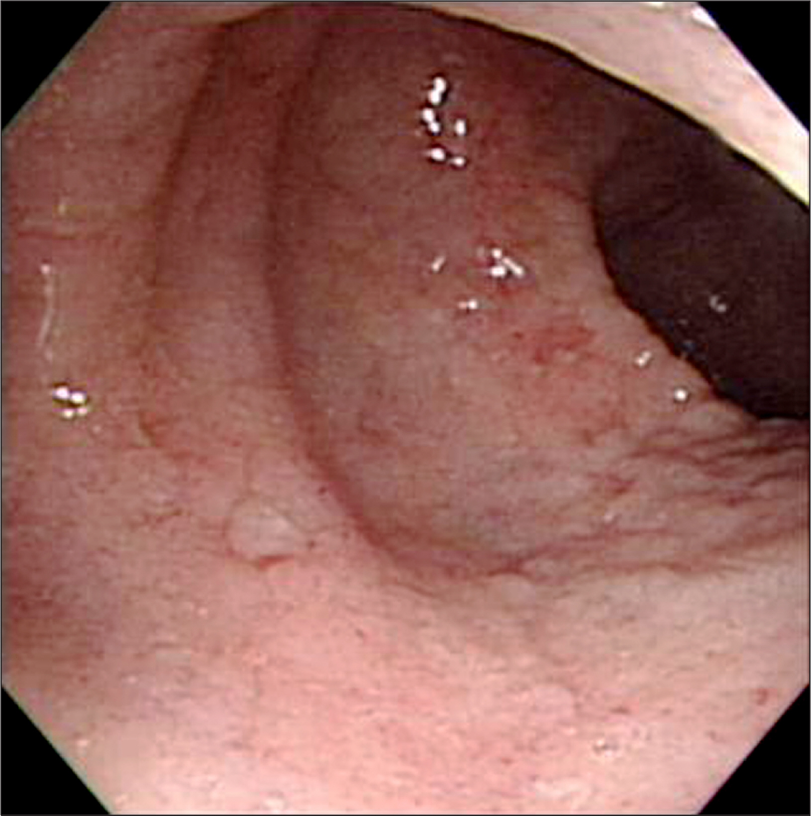
Fig. 1. Colonoscopic finding. Colonoscopy showed shallow ulceration at the terminal ileum.
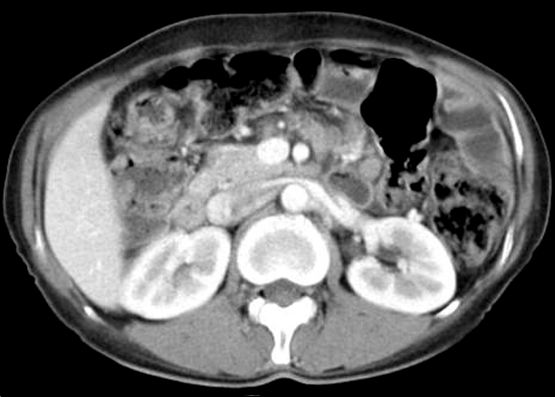
Fig. 2. Abdominal CT finding. Abdominal CT showed multiple conglomerated lymphadenopathies in the mesentery.
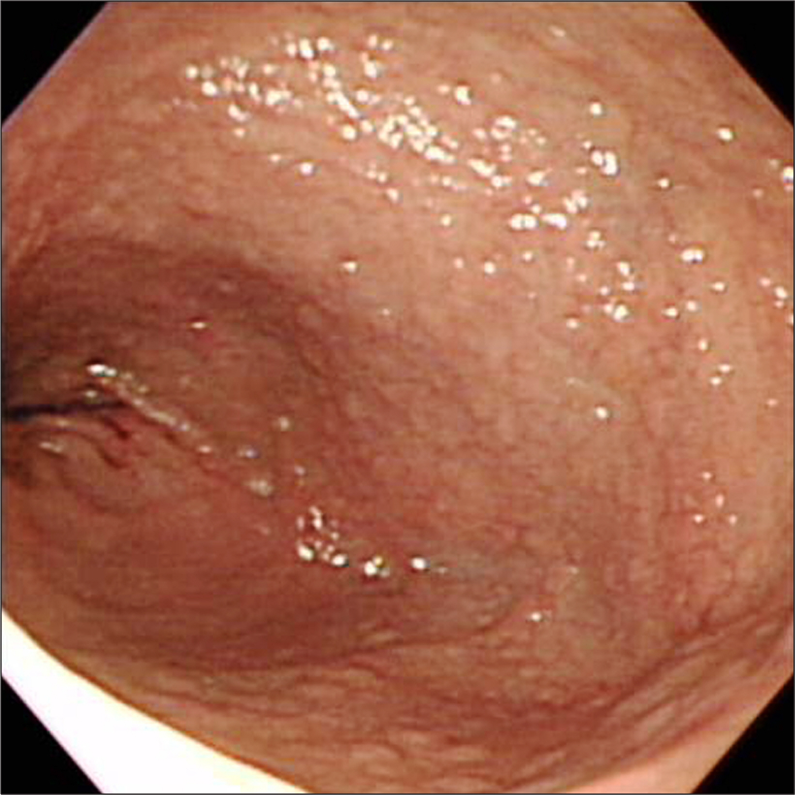
Fig. 3. Endoscopic finding. It showed diffuse micronodularity at the duodenal 2nd portion.

Fig. 4. Microscopic findings of the duodenal 2nd portion. (A) Villous atrophy, crypt hyperplasia in a biopsy of duodenum 2nd portion (H&E, ×100).(B) Increase in the intraepithelial lymphocytes was noted (H&E, ×400).

Fig. 5. Capsule endoscopic finding. It showed blunted villi with focal atrophic change at the jejunum.
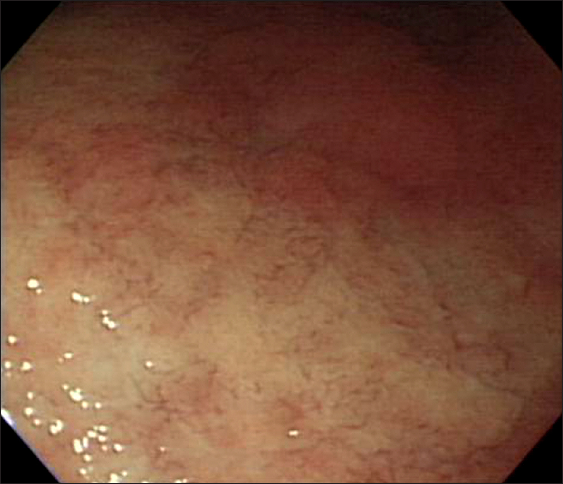
Fig. 6. Enteroscopic finding. It showed diffuse atrophic villous mucosal change with blunting of villi at the jejunum.

Fig. 7. Microscopic findings of the jejunum. (A) It revealed marked chronic inflmmation with lymphoid aggregation and blunting of villi (H&E, ×100). (B) It revealed lymphoid aggregation and villous atrophy (H&E, ×400).
Cited by 3 articles
-
Non-celiac Gluten Sensitivity
Ra Ri Cha, Hyun Jin Kim
Korean J Gastroenterol. 2020;75(1):11-16. doi: 10.4166/kjg.2020.75.1.11.A case of celiac disease with neurologic manifestations misdiagnosed as amyotrophic lateral sclerosis
Hyoju Ham, Bo-In Lee, Hyun Jin Oh, Se Hwan Park, Jin Su Kim, Jae Myung Park, Young Seok Cho, Myung-Gyu Choi
Intest Res. 2017;15(4):540-542. doi: 10.5217/ir.2017.15.4.540.Current status and future perspectives of capsule endoscopy
Hyun Joo Song, Ki-Nam Shim
Intest Res. 2016;14(1):21-29. doi: 10.5217/ir.2016.14.1.21.
Reference
-
References
1. Mäki M, Mustalahti K, Kokkonen J, et al. Prevalence of celiac disease among children in Finland. N Engl J Med. 2003; 348:2517–2524.
Article2. Rubio-Tapia A, Ludvigsson JF, Brantner TL, Murray JA, Everhart JE. The prevalence of celiac disease in the United States. Am J Gastroenterol. 2012; 107:1538–1544.
Article3. Corazza GR, Di Sario A, Cecchetti L, et al. Bone mass and metabolism in patients with celiac disease. Gastroenterology. 1995; 109:122–128.
Article4. Kemppainen T, Kröger H, Janatuinen E, et al. Osteoporosis in adult patients with celiac disease. Bone. 1999; 24:249–255.
Article5. James SP. National Institutes of Health Consensus Development Conference Statement on Celiac Disease, June 28–30, 2004. Gastroenterology. 2005; 128(4 Suppl 1):S1–S9.
Article6. Rostom A, Murray JA, Kagnoff MF. American Gastroenterological Association (AGA) Institute technical review on the diagnosis and management of celiac disease. Gastroenterology. 2006; 131:1981–2002.
Article7. Armstrong MJ, Hegade VS, Robins G. Advances in coeliac disease. Curr Opin Gastroenterol. 2012; 28:104–112.
Article8. Cummins AG, Roberts-Thomson IC. Prevalence of celiac disease in the Asia-Pacific region. J Gastroenterol Hepatol. 2009; 24:1347–1351.
Article
- Full Text Links
-
- Actions
-
Cited
- CITED
-
- Close
- Share
-
- Similar articles
-
- Malignant Histiocytic Lymphoma Associated with Celiac Disease: A Case Report
- Association between Celiac Disease and Intussusceptions in Children: Two Case Reports and Literature Review
- Rhabdomyolysis in Celiac Disease
- Spontaneous Isolated Dissection of the Celiac Artery: a Case Report
- A case of enteropathy-type intestinal T-cell lymphoma, confused with celiac disease
