Mixed Echoic Thyroid Nodules on Ultrasound: Approach to Management
- Affiliations
-
- 1Department of Radiology, Kyung Hee University Hospital, College of Medicine, Kyung Hee University, Seoul, Korea.
- 2Department of Radiology, CHA Bundang Medical Center, CHA University College of Medicine, Seongnam, Korea.
- 3Department of Radiology, Research Institute of Radiological Science, Yonsei University College of Medicine, Seoul, Korea. docjin@yuhs.ac
- KMID: 1716883
- DOI: http://doi.org/10.3349/ymj.2012.53.4.812
Abstract
- PURPOSE
To evaluate malignancy risk according to ultrasound (US) features and size change on follow-up US in mixed echoic thyroid nodules and to suggest management guidelines thereof.
MATERIALS AND METHODS
Among patients who underwent US-guided fine needle aspiration biopsy, 316 mixed echoic nodules in 303 patients were included after excluding the patients with pure solid or cystic nodules or without further cytopathologic evaluation. We evaluated malignancy risk according to US features and changes in size and shape on follow-up US.
RESULTS
The malignancy rate was 31.6% (6 of 19) for nodules with suspicious US features and 2.7% (8 of 297) for nodules without suspicious US features (p<0.001). Among 265 nodules with no suspicious US features and initial benign cytology, 15 nodules with suspicious US change and decreased size, 25 nodules with no suspicious US change and increased size, and 225 nodules with no suspicious US change and no change in size were observed on follow-up USs. The malignancy risk thereof was 0%, 0% and 0.4%, respectively (p=1.000).
CONCLUSION
Mixed echoic nodules with no suspicious US features and benign cytology can be followed up using US, as they revealed very low malignancy rates, even if they showed growth on follow-up US.
MeSH Terms
Figure
-
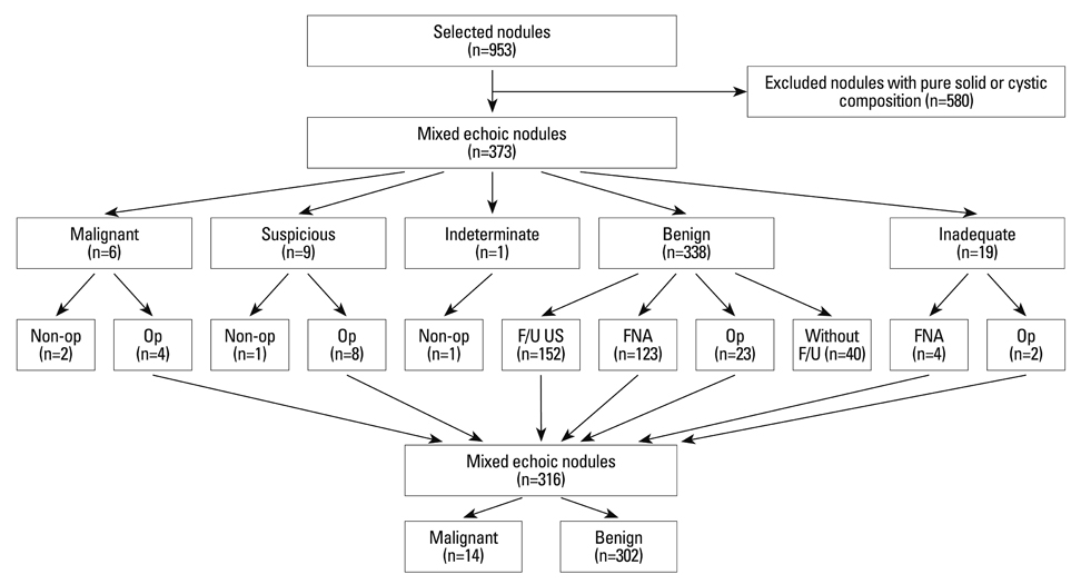
Fig. 1 Diagram of the study population. non-op, without operation; Op, with operation; FNA, fine needle aspiration.
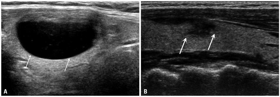
Fig. 2 A 51-year-old woman with a thyroid nodule in the right thyroid on US. Initial FNAB revealed the nodule to be benign on cytology, and follow-up FNAB after 1.5 year showed the same cytologic result. (A) Initial US shows a mixed echoic nodule (>50% cystic) (arrows). (B) On follow-up US, this nodule showed decreased size and suspicious US changes including hypoechoic and microlobulated margins and taller-than wide shape (arrows). US, ultrasound; FNAB, fine needle aspiration biopsy.
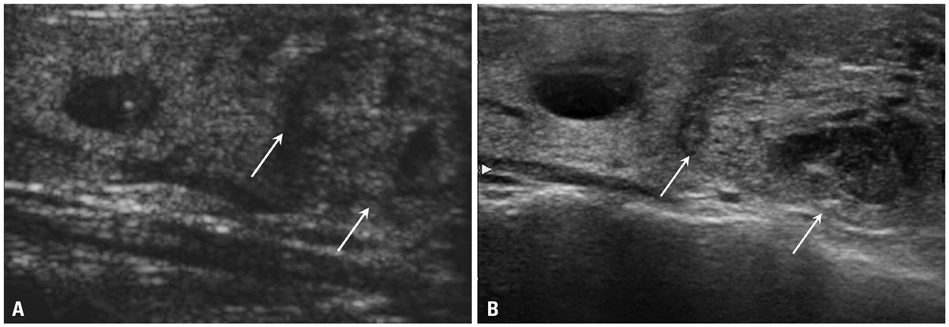
Fig. 3 A 56-year-old woman with a thyroid nodule in the right thyroid on US. Initial FNAB revealed the nodule to be benign on cytology, and follow-up FNAB after 3 year showed the same cytologic result. (A) Initial US shows a mixed echoic nodule (< 50% cystic) (arrows). (B) On follow-up US, this nodule showed increased size without change in shape (arrows). US, ultrasound; FNAB, fine needle aspiration biopsy.
Cited by 3 articles
-
Anaplastic Thyroid Cancer: Ultrasonographic Findings and the Role of Ultrasonography-Guided Fine Needle Aspiration Biopsy
Hee Jung Suh, Hee Jung Moon, Jin Young Kwak, Ji Soo Choi, Eun-Kyung Kim
Yonsei Med J. 2013;54(6):1400-1406. doi: 10.3349/ymj.2013.54.6.1400.Diagnostic Role of Conventional Ultrasonography and Shearwave Elastography in Asymptomatic Patients with Diffuse Thyroid Disease: Initial Experience with 57 Patients
Injoong Kim, Eun-Kyung Kim, Jung Hyun Yoon, Kyung Hwa Han, Eun Ju Son, Hee Jung Moon, Jin Young Kwak
Yonsei Med J. 2014;55(1):247-253. doi: 10.3349/ymj.2014.55.1.247.Pathologic Spectrum of Lymphocytic Infiltration and Recurrence of Papillary Thyroid Carcinoma
Hyun Gi Kim, Eun-Kyung Kim, Kyung Hwa Han, Hyunki Kim, Jin Young Kwak
Yonsei Med J. 2014;55(4):879-885. doi: 10.3349/ymj.2014.55.4.879.
Reference
-
1. Mazzaferri EL. Management of a solitary thyroid nodule. N Engl J Med. 1993. 328:553–559.
Article2. Choi KU, Kim JY, Park DY, Lee CH, Sol MY, Han KT, et al. Recommendations for the management of cystic thyroid nodules. ANZ J Surg. 2005. 75:537–541.
Article3. de los Santos ET, Keyhani-Rofagha S, Cunningham JJ, Mazzaferri EL. Cystic thyroid nodules. The dilemma of malignant lesions. Arch Intern Med. 1990. 150:1422–1427.
Article4. McHenry CR, Slusarczyk SJ, Khiyami A. Recommendations for management of cystic thyroid disease. Surgery. 1999. 126:1167–1171.
Article5. Meko JB, Norton JA. Large cystic/solid thyroid nodules: a potential false-negative fine-needle aspiration. Surgery. 1995. 118:996–1003.
Article6. Bellantone R, Lombardi CP, Raffaelli M, Traini E, De Crea C, Rossi ED, et al. Management of cystic or predominantly cystic thyroid nodules: the role of ultrasound-guided fine-needle aspiration biopsy. Thyroid. 2004. 14:43–47.
Article7. Nam-Goong IS, Kim HY, Gong G, Lee HK, Hong SJ, Kim WB, et al. Ultrasonography-guided fine-needle aspiration of thyroid incidentaloma: correlation with pathological findings. Clin Endocrinol (Oxf). 2004. 60:21–28.
Article8. Lee MJ, Kim EK, Kwak JY, Kim MJ. Partially cystic thyroid nodules on ultrasound: probability of malignancy and sonographic differentiation. Thyroid. 2009. 19:341–346.
Article9. Crile G Jr. Treatment of thyroid cysts by aspiration. Surgery. 1966. 59:210–212.10. Sarda AK, Bal S, Dutta Gupta S, Kapur MM. Diagnosis and treatment of cystic disease of the thyroid by aspiration. Surgery. 1988. 103:593–596.11. Frates MC, Benson CB, Doubilet PM, Kunreuther E, Contreras M, Cibas ES, et al. Prevalence and distribution of carcinoma in patients with solitary and multiple thyroid nodules on sonography. J Clin Endocrinol Metab. 2006. 91:3411–3417.
Article12. Alexander EK, Heering JP, Benson CB, Frates MC, Doubilet PM, Cibas ES, et al. Assessment of nondiagnostic ultrasound-guided fine needle aspirations of thyroid nodules. J Clin Endocrinol Metab. 2002. 87:4924–4927.
Article13. Braga M, Cavalcanti TC, Collaço LM, Graf H. Efficacy of ultrasound-guided fine-needle aspiration biopsy in the diagnosis of complex thyroid nodules. J Clin Endocrinol Metab. 2001. 86:4089–4091.
Article14. Kuma K, Matsuzuka F, Yokozawa T, Miyauchi A, Sugawara M. Fate of untreated benign thyroid nodules: results of long-term follow-up. World J Surg. 1994. 18:495–498.
Article15. Singer PA, Cooper DS, Daniels GH, Ladenson PW, Greenspan FS, Levy EG, et al. American Thyroid Association. Treatment guidelines for patients with thyroid nodules and well-differentiated thyroid cancer. Arch Intern Med. 1996. 156:2165–2172.
Article16. American Thyroid Association (ATA) Guidelines Taskforce on Thyroid Nodules and Differentiated Thyroid Cancer. Cooper DS, Doherty GM, Haugen BR, Kloos RT, Lee SL, et al. Revised American Thyroid Association management guidelines for patients with thyroid nodules and differentiated thyroid cancer. Thyroid. 2009. 19:1167–1214.
Article17. Frates MC, Benson CB, Charboneau JW, Cibas ES, Clark OH, Coleman BG, et al. Management of thyroid nodules detected at US: Society of Radiologists in Ultrasound consensus conference statement. Radiology. 2005. 237:794–800.
Article18. Gharib H, Papini E, Paschke R, Duick DS, Valcavi R, Hegedüs L, et al. American Association of Clinical Endocrinologists, Associazione Medici Endocrinologi, and European Thyroid Association Medical Guidelines for Clinical Practice for the Diagnosis and Management of Thyroid Nodules. Endocr Pract. 2010. 16:Suppl 1. 1–43.
Article19. Koo JH, Shin JH, Han BK, Ko EY, Kang SS. Cystic thyroid nodules after aspiration mimicking malignancy: sonographic characteristics. J Ultrasound Med. 2010. 29:1415–1421.20. Kim EK, Park CS, Chung WY, Oh KK, Kim DI, Lee JT, et al. New sonographic criteria for recommending fine-needle aspiration biopsy of nonpalpable solid nodules of the thyroid. AJR Am J Roentgenol. 2002. 178:687–691.
Article21. Alexander EK, Hurwitz S, Heering JP, Benson CB, Frates MC, Doubilet PM, et al. Natural history of benign solid and cystic thyroid nodules. Ann Intern Med. 2003. 138:315–318.
Article22. Müller N, Cooperberg PL, Suen KC, Thorson SC. Needle aspiration biopsy in cystic papillary carcinoma of the thyroid. AJR Am J Roentgenol. 1985. 144:251–253.
Article23. Gharib H, Papini E, Valcavi R, Baskin HJ, Crescenzi A, Dottorini ME, et al. American Association of Clinical Endocrinologists and Associazione Medici Endocrinologi medical guidelines for clinical practice for the diagnosis and management of thyroid nodules. Endocr Pract. 2006. 12:63–102.24. Kwak JY, Kim EK, Kim HJ, Kim MJ, Son EJ, Moon HJ. How to combine ultrasound and cytological information in decision making about thyroid nodules. Eur Radiol. 2009. 19:1923–1931.
Article25. Kim DW, Lee EJ, In HS, Kim SJ. Sonographic differentiation of partially cystic thyroid nodules: a prospective study. AJNR Am J Neuroradiol. 2010. 31:1961–1966.
Article26. Cappelli C, Castellano M, Pirola I, Gandossi E, De Martino E, Cumetti D, et al. Thyroid nodule shape suggests malignancy. Eur J Endocrinol. 2006. 155:27–31.
Article27. Iannuccilli JD, Cronan JJ, Monchik JM. Risk for malignancy of thyroid nodules as assessed by sonographic criteria: the need for biopsy. J Ultrasound Med. 2004. 23:1455–1464.
Article28. Moon WJ, Baek JH, Jung SL, Kim DW, Kim EK, Kim JY, et al. Ultrasonography and the ultrasound-based management of thyroid nodules: consensus statement and recommendations. Korean J Radiol. 2011. 12:1–14.
Article29. Moon WJ, Jung SL, Lee JH, Na DG, Baek JH, Lee YH, et al. Benign and malignant thyroid nodules: US differentiation--multicenter retrospective study. Radiology. 2008. 247:762–770.
Article30. Papini E, Guglielmi R, Bianchini A, Crescenzi A, Taccogna S, Nardi F, et al. Risk of malignancy in nonpalpable thyroid nodules: predictive value of ultrasound and color-Doppler features. J Clin Endocrinol Metab. 2002. 87:1941–1946.
Article31. Tae HJ, Lim DJ, Baek KH, Park WC, Lee YS, Choi JE, et al. Diagnostic value of ultrasonography to distinguish between benign and malignant lesions in the management of thyroid nodules. Thyroid. 2007. 17:461–466.
Article32. Alexander EK, Marqusee E, Orcutt J, Benson CB, Frates MC, Doubilet PM, et al. Thyroid nodule shape and prediction of malignancy. Thyroid. 2004. 14:953–958.
Article33. Lee MJ, Hong SW, Chung WY, Kwak JY, Kim MJ, Kim EK. Cytological results of ultrasound-guided fine-needle aspiration cytology for thyroid nodules: emphasis on correlation with sonographic findings. Yonsei Med J. 2011. 52:838–844.
Article34. Cappelli C, Castellano M, Pirola I, Cumetti D, Agosti B, Gandossi E, et al. The predictive value of ultrasound findings in the management of thyroid nodules. QJM. 2007. 100:29–35.
Article35. Ito Y, Kobayashi K, Tomoda C, Uruno T, Takamura Y, Miya A, et al. Ill-defined edge on ultrasonographic examination can be a marker of aggressive characteristic of papillary thyroid microcarcinoma. World J Surg. 2005. 29:1007–1011.
Article36. Kang HW, No JH, Chung JH, Min YK, Lee MS, Lee MK, et al. Prevalence, clinical and ultrasonographic characteristics of thyroid incidentalomas. Thyroid. 2004. 14:29–33.
Article37. Koike E, Noguchi S, Yamashita H, Murakami T, Ohshima A, Kawamoto H, et al. Ultrasonographic characteristics of thyroid nodules: prediction of malignancy. Arch Surg. 2001. 136:334–337.
Article38. Peccin S, de Castsro JA, Furlanetto TW, Furtado AP, Brasil BA, Czepielewski MA. Ultrasonography: is it useful in the diagnosis of cancer in thyroid nodules? J Endocrinol Invest. 2002. 25:39–43.
Article39. Cappelli C, Pirola I, Cumetti D, Micheletti L, Tironi A, Gandossi E, et al. Is the anteroposterior and transverse diameter ratio of nonpalpable thyroid nodules a sonographic criteria for recommending fine-needle aspiration cytology? Clin Endocrinol (Oxf). 2005. 63:689–693.
Article40. Chan BK, Desser TS, McDougall IR, Weigel RJ, Jeffrey RB Jr. Common and uncommon sonographic features of papillary thyroid carcinoma. J Ultrasound Med. 2003. 22:1083–1090.
Article41. Khoo ML, Asa SL, Witterick IJ, Freeman JL. Thyroid calcification and its association with thyroid carcinoma. Head Neck. 2002. 24:651–655.
Article42. Rago T, Vitti P, Chiovato L, Mazzeo S, De Liperi A, Miccoli P, et al. Role of conventional ultrasonography and color flow-doppler sonography in predicting malignancy in 'cold' thyroid nodules. Eur J Endocrinol. 1998. 138:41–46.
Article43. Frates MC, Benson CB, Doubilet PM, Cibas ES, Marqusee E. Can color Doppler sonography aid in the prediction of malignancy of thyroid nodules? J Ultrasound Med. 2003. 22:127–131.
Article44. Hatabu H, Kasagi K, Yamamoto K, Iida Y, Misaki T, Hidaka A, et al. Cystic papillary carcinoma of the thyroid gland: a new sonographic sign. Clin Radiol. 1991. 43:121–124.
Article45. Kwak JY, Koo H, Youk JH, Kim MJ, Moon HJ, Son EJ, et al. Value of US correlation of a thyroid nodule with initially benign cytologic results. Radiology. 2010. 254:292–300.
Article46. Yoon JH, Moon HJ, Kim EK, Kwak JY. Inadequate cytology in thyroid nodules: should we repeat aspiration or follow-up? Ann Surg Oncol. 2011. 18:1282–1289.
Article
- Full Text Links
-
- Actions
-
Cited
- CITED
-
- Close
- Share
-
- Similar articles
-
- Elastography of the Thyroid Glands
- Ultrasound (US)-Guided Ablation of Thyroid Nodules
- Detection and Management of Thyroid incidentalomas
- Diagnosis of Parathyroid Adenoma Detected during Thyroid Ultrasound: The Role of Parathormone Measurement in Fine-Needle Aspiration Washout
- Updated guidelines for the diagnosis and management of thyroid nodules
