Lab Anim Res.
2013 Dec;29(4):183-189. 10.5625/lar.2013.29.4.183.
Is sheep lumbar spine a suitable alternative model for human spinal researches? Morphometrical comparison study
- Affiliations
-
- 1Large Animal Clinic for Surgery, Faculty of Veterinary Medicine, University of Leipzig, Leipzig, Germany. mahmoud.mageed@hotmail.com
- 2Department of Surgery and Anaesthesia, Faculty of Veterinary Medicine, University of Khartoum, Khartoum North, Sudan.
- 3Microsurgery and Animal Models Core, Translational Centre for Regenerative Medicine, University of Leipzig, Leipzig, Germany.
- 4Department of Neurosurgery, BG Hospital Bergmannstrost, Halle, Germany.
- KMID: 1716212
- DOI: http://doi.org/10.5625/lar.2013.29.4.183
Abstract
- Sheep are commonly used as a model for human spinal orthopaedic research due to their similarity in morphological and biomechanical features. This study aimed to document the volumes of vertebral bodies and compare the generated results as well as morphometry of the sheep lumbar spine to human published data. For this purpose, computed tomography scans were carried out on five adult Merino sheep under general anaesthesia. Transverse 5 mm thick images were acquired from L1 to L6 using a multi-detector-row helical CT scanner. Volume measurements were performed with dedicated software. Four spinal indices and Pavlov's ratio were calculated. Thereafter, the generated data were compared to published literature on humans. The mean vertebral body volume showed an increase towards the caudal vertebrae, but there were no significant differences between the vertebral levels (P>0.05). Compared to humans, sheep vertebral body volumes were 48.6% smaller. The comparison of absolute values between both species revealed that sheep had smaller, longer and narrower vertebral bodies, thinner intervertebral discs, narrower spinal canal, longer transverse processes, shorter dorsal spinous processes and narrower, higher pedicles with more lateral angulations. The comparison of the spinal indices showed a good similarity to human in terms of the vertebral endplates and spinal canal. The results of this study may be helpful for using the sheep as a model for human orthopaedic spinal research if anatomical differences are taken into account.
MeSH Terms
Figure
-
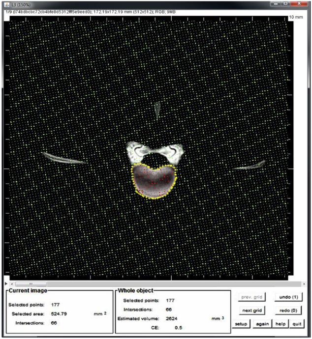
Figure 1 Estimation the vertebral body volume using ImageJ. The vertebral body was outlined (yellow stars), subsequently the Volumest plugin calculates automatically the volume from the area (red stars) and the slice thickness of each image. Finally, these values per image were added up to calculate the volume.
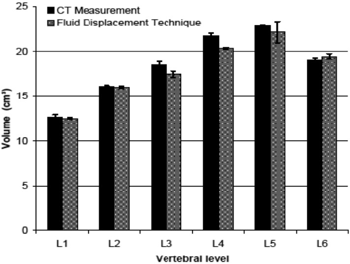
Figure 2 The means of the vertebral body volumes (cm3) of the sheep using imageJ software compared to real volume of the same vertebrae, which was measured using fluid displacement technique.
Reference
-
1. Putz RL, Müller-Gerbl M. The vertebral column--a phylogenetic failure? A theory explaining the function and vulnerability of the human spine. Clin Anat. 1996; 9(3):205–212. PMID: 8740482.
Article2. Mann NH, Brown MD, Enger I. Statistical diagnosis of lumbar spine disorders using computerized patient pain drawings. Comput Biol Med. 1991; 21(6):383–397. PMID: 1838724.
Article3. Ashman RB, Bechtold JE, Edwards WT, Johnston CE, McAfee PC, Tencer AF. In vitro spinal arthrodesis implant mechanical testing protocols. J Spinal Disord. 1989; 2(4):274–281. PMID: 2520086.
Article4. Wall EJ, Bylski-Austrow DI, Shelton FS, Crawford AH, Kolata RJ, Baum DS. Endoscopic discectomy increases thoracic spine flexibility as effectively as open discectomy. A mechanical study in a porcine model. Spine. 1998; 23(1):9–15. PMID: 9460146.5. Reid JE, Meakin JR, Robins SP, Skakle JM, Hukins DW. Sheep lumbar intervertebral discs as models for human discs. Clin Biomech (Bristol, Avon). 2002; 17(4):312–314.
Article6. Gurr KR, McAfee PC, Shih CM. Biomechanical analysis of anterior and posterior instrumentation systems after corpectomy. A calf-spine model. J Bone Joint Surg Am. 1988; 70(8):1182–1191. PMID: 3417703.
Article7. Ganey T, Libera J, Moos V, Alasevic O, Fritsch KG, Meisel HJ, Hutton WC. Disc chondrocyte transplantation in a canine model: a treatment for degenerated or damaged intervertebral disc. Spine. 2003; 28(23):2609–2620. PMID: 14652478.
Article8. Newman E, Turner AS, Wark JD. The potential of sheep for the study of osteopenia: current status and comparison with other animal models. Bone. 1995; 16(4):277S–284S. PMID: 7626315.
Article9. Nunamaker D. Experimental models of fracture repair. Clin Orthop Relat Res. 1998; 355:S56–S65. PMID: 9917626.
Article10. Bergmann G, Graichen F, Rohlmann A. Hip joint forces in sheep. J Biomech. 1999; 32(8):769–777. PMID: 10433418.
Article11. Egermann M, Goldhahn J, Schneider E. Animal models for fracture treatment in osteoporosis. Osteoporos Int. 2005; 16(Suppl 2):S129–S138. PMID: 15750681.
Article12. Turner AS. Experiences with sheep as an animal model for shoulder surgery: strengths and shortcomings. J Shoulder Elbow Surg. 2007; 16(5):S158–S163. PMID: 17507248.
Article13. Ahlgren BD, Lui W, Herkowitz HN, Panjabi MM, Guiboux JP. Effect of anular repair on the healing strength of the intervertebral disc: a sheep model. Spine. 2000; 25(17):2165–2170. PMID: 10973397.14. Moore RJ, Osti OL, Vernon-Roberts B, Fraser RD. Changes in endplate vascularity after an outer anulus tear in the sheep. Spine. 1992; 17(8):874–878. PMID: 1523489.
Article15. Limthongkul W, Karaikovic EE, Savage JW, Markovic A. Volumetric analysis of thoracic and lumbar vertebral bodies. Spine J. 2010; 10(2):153–158. PMID: 20142072.
Article16. Odaci E, Sahin B, Sonmez OF, Kaplan S, Bas O, Bilgic S, Bek Y, Ergür H. Rapid estimation of the vertebral body volume: a combination of the Cavalieri principle and computed tomography images. Eur J Radiol. 2003; 48(3):316–326. PMID: 14652153.
Article17. Merzin M. Applying stereological method in radiology: Volume measurement (Bachelor's thesis). Tartu: University of Tartu;2008.18. Mageed M, Berner D, Jülke H, Hohaus C, Brehm W, Gerlach K. Morphometrical dimensions of the sheep thoracolumbar vertebrae as seen on digitised CT images. Lab Anim Res. 2013; 29(3):138–147. PMID: 24106508.
Article19. Abuzayed B, Tutunculer B, Kucukyuruk B, Tuzgen S. Anatomic basis of anterior and posterior instrumentation of the spine: morphometric study. Surg Radiol Anat. 2010; 32(1):75–85. PMID: 19696959.
Article20. Kumar N, Kukreti S, Ishaque M, Mulholland R. Anatomy of deer spine and its comparison to the human spine. Anat Rec. 2000; 260(2):189–203. PMID: 10993955.
Article21. Wolf A, Shoham M, Michael S, Moshe R. Morphometric study of the human lumbar spine for operation-workspace specifications. Spine. 2001; 26(22):2472–2477. PMID: 11707713.
Article22. Pavlov H, Torg JS, Robie B, Jahre C. Cervical spinal stenosis: determination with vertebral body ratio method. Radiology. 1987; 164(3):771–775. PMID: 3615879.
Article23. Beers GJ, Carter AP, Leiter BE, Tilak SP, Shah RR. Interobserver discrepancies in distance measurements from lumbar spine CT scans. AJR Am J Roentgenol. 1985; 144(2):395–398. PMID: 3871289.
Article24. Wilke HJ, Kettler A, Wenger KH, Claes LE. Anatomy of the sheep spine and its comparison to the human spine. Anat Rec. 1997; 247(4):542–555. PMID: 9096794.
Article25. Pluijm SM, Tromp AM, Smit JH, Deeg DJ, Lips P. Consequences of vertebral deformities in older men and women. J Bone Miner Res. 2000; 15(8):1564–1572. PMID: 10934655.
Article26. Busscher I, Ploegmakers JJ, Verkerke GJ, Veldhuizen AG. Comparative anatomical dimensions of the complete human and porcine spine. Eur Spine J. 2010; 19(7):1104–1114. PMID: 20186441.
Article27. Wilke HJ, Kettler A, Claes LE. Are sheep spines a valid biomechanical model for human spines? Spine. 1997; 22(20):2365–2374. PMID: 9355217.
Article28. Masharawi Y, Salame K, Mirovsky Y, Peleg S, Dar G, Steinberg N, Hershkovitz I. Vertebral body shape variation in the thoracic and lumbar spine: characterization of its asymmetry and wedging. Clin Anat. 2008; 21(1):46–54. PMID: 17729333.
Article
- Full Text Links
-
- Actions
-
Cited
- CITED
-
- Close
- Share
-
- Similar articles
-
- Morphometrical dimensions of the sheep thoracolumbar vertebrae as seen on digitised CT images
- Spinal anesthesia in elective lumbar spinal surgery
- Regional Anesthesia for Lumbar Spine Surgery: Can It Be a Standard in the Future?
- Long-Term Effects of Segmental Lumbar Spinal Fusion on Adjacent Healthy Discs: A Finite Element Study
- Measurement of the Oblique Diameter of the Lumbar Spinal Canal in Korean Army-aged Group by Echographic Method
