Portable Document Format File Showing the Surface Models of Cadaver Whole Body
- Affiliations
-
- 1Department of Anatomy, Ajou University School of Medicine, Suwon, Korea.
- 2Department of Anatomy, Dongguk University College of Medicine, Gyeongju, Korea.
- 3Korea Institute of Science and Technology Information, Daejeon, Korea.
- 4Department of Orthopedics, Chosun University Hospital, Gwangju, Korea.
- 5Graduate School of Information and Communication, Ajou University, Suwon, Korea. crjang@ajou.ac.kr
- KMID: 1714194
- DOI: http://doi.org/10.3346/jkms.2012.27.8.849
Abstract
- In the Visible Korean project, 642 three-dimensional (3D) surface models have been built from the sectioned images of a male cadaver. It was recently discovered that popular PDF file enables users to approach the numerous surface models conveniently on Adobe Reader. Purpose of this study was to present a PDF file including systematized surface models of human body as the beneficial contents. To achieve the purpose, fitting software packages were employed in accordance with the procedures. Two-dimensional (2D) surface models including the original sectioned images were embedded into the 3D surface models. The surface models were categorized into systems and then groups. The adjusted surface models were inserted to a PDF file, where relevant multimedia data were added. The finalized PDF file containing comprehensive data of a whole body could be explored in varying manners. The PDF file, downloadable freely from the homepage (http://anatomy.co.kr), is expected to be used as a satisfactory self-learning tool of anatomy. Raw data of the surface models can be extracted from the PDF file and employed for various simulations for clinical practice. The technique to organize the surface models will be applied to manufacture of other PDF files containing various multimedia contents.
Keyword
MeSH Terms
Figure
-
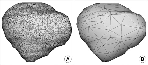
Fig. 1 The surface model of the patella with its mesh. (A) Full data. (B) Reduced data.

Fig. 2 Sectioned images with labels, embedded on the surface models.
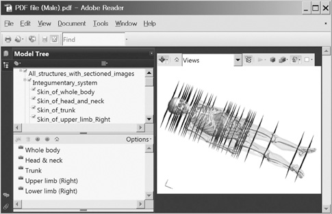
Fig. 3 Feature of PDF file, composed of the model tree window (left top), bookmark window (left bottom), and surface models (right). The surface models are colored with the skin made semitransparent.
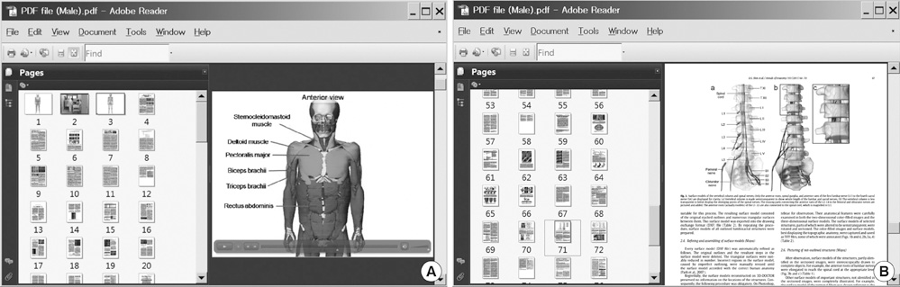
Fig. 4 Movie and paper introducing the surface models. (A) In the PDF files, the first and second movie is located on page 2 and 3, respectively. (B) The related articles are situated on the remnant pages.

Fig. 5 Combination of the surface models showing the anatomical knowledge that esophagus is compressed by three structures: the arch of aorta, left main bronchus, and diaphragm.
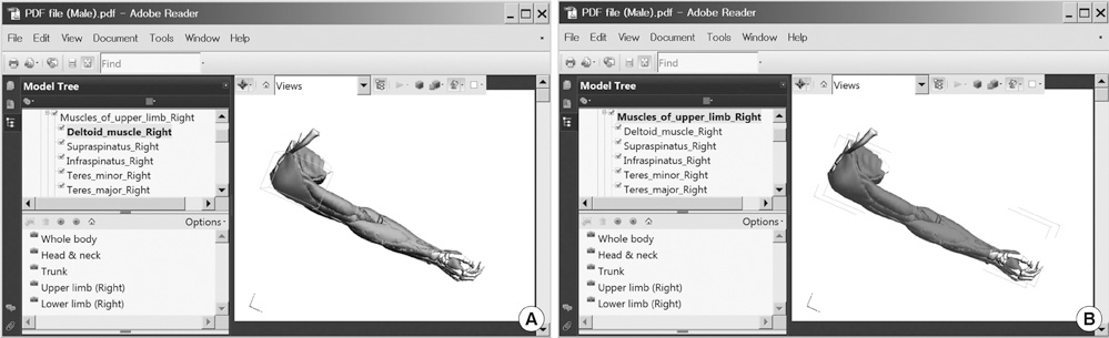
Fig. 6 Zoomed-in right upper limb region by selection of the bookmark 'Upper limb (Right).' (A) Highlighting of the surface model 'deltoid muscle' coincides with highlighting of the text 'deltoid muscle'. (B) Multiple clicks induce the spotlight of the muscular system including 'deltoid'.

Fig. 7 Sectioning of the surface models of digestive tract. The horizontal (A), coronal (B), and sagittal (C) planes.
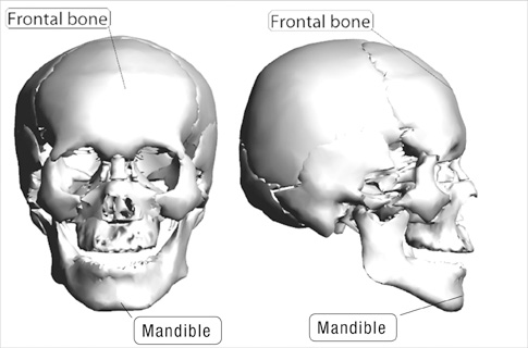
Fig. 8 Stereoscopic labeling on the surface models of frontal bone and mandible.
Cited by 7 articles
-
Portable Document Format File Containing the Schematics and Operable Surface Models of the Head Structures
Beom Sun Chung, Min Suk Chung, Jin Seo Park
J Korean Med Sci. 2020;35(27):e212. doi: 10.3346/jkms.2020.35.e212.Three Software Tools for Viewing Sectional Planes, Volume Models, and Surface Models of a Cadaver Hand
Beom Sun Chung, Min Suk Chung, Byeong-Seok Shin, Koojoo Kwon
J Korean Med Sci. 2018;33(8):. doi: 10.3346/jkms.2018.33.e64.Rise of the Visible Monkey: Sectioned Images of Rhesus Monkey
Beom Sun Chung, Chang-Yeop Jeon, Jae-Won Huh, Kang-Jin Jeong, Donghwan Har, Kyu-Sung Kwack, Jin Seo Park
J Korean Med Sci. 2019;34(8):. doi: 10.3346/jkms.2019.34.e66.New Viewpoint of Surface Anatomy Using the Curved Sectional Planes of a Male Cadaver
Koojoo Kwon, Byeong-Seok Shin, Min Suk Chung, Beom Sun Chung
J Korean Med Sci. 2019;34(3):. doi: 10.3346/jkms.2019.34.e15.Effects of Reading a Free Electronic Book on Regional Anatomy with Schematics and Mnemonics on Student Learning
Beom Sun Chung, Ki Seok Koh, Chang-Seok Oh, Jin Seo Park, Jae-Ho Lee, Min Suk Chung
J Korean Med Sci. 2020;35(6):. doi: 10.3346/jkms.2020.35.e42.Homepage to distribute the anatomy learning contents including Visible Korean products, comics, and books
Beom Sun Chung, Min Suk Chung
Anat Cell Biol. 2018;51(1):7-13. doi: 10.5115/acb.2018.51.1.7.A Guide Book for Children and Adolescents to Experience Anatomy and Clinics
Bum Sun Chung, Seong-bae Koh, Eun-mi Park, Mi-Geum Song, Seo-eun Lee, Tae-ho Jeon, Sook-kyoung Cho, Min Suk Chung
Korean J Phys Anthropol. 2015;28(2):79-85. doi: 10.11637/kjpa.2015.28.2.79.
Reference
-
1. Park JS, Chung MS, Hwang SB, Lee YS, Har DH, Park HS. Visible Korean Human. Improved serially sectioned images of the entire body. IEEE Trans Med Imaging. 2005. 24:352–360.2. Park JS, Chung MS, Hwang SB, Lee YS, Har DH, Park HS. Technical report on semiautomatic segmentation by using the Adobe Photoshop. J Digit Imaging. 2005. 18:333–343.3. Shin DS, Park JS, Park HS, Hwang SB, Chung MS. Outlining of the detailed structures in sectioned images from Visible Korean. Surg Radiol Anat. 2012. 34:235–247.4. Shin DS, Chung MS, Lee JW, Park JS, Chung J, Lee SB, Lee SH. Advanced surface reconstruction technique to build detailed surface models of liver and neighboring structures from the Visible Korean Human. J Korean Med Sci. 2009. 24:375–383.5. Shin DS, Park JS, Lee SB, Lee SH, Chung J, Chung MS. Surface model of the gastrointestinal tract constructed from the Visible Korean. Clin Anat. 2009. 22:601–609.6. Jang HG, Chung MS, Shin DS, Park SK, Cheon KS, Park JS. Segmentation and surface reconstruction of the detailed ear structures, identified in sectioned images. Anat Rec (Hoboken). 2011. 294:559–564.7. Shin DS, Chung MS, Park JS, Park HS, Lee SB, Lee SH, Choi HN, Riemer M, Handels H, Lee JE, et al. Three-dimensional surface models of detailed lumbosacral structures reconstructed from the Visible Korean. Ann Anat. 2011. 193:64–70.8. Shin DS, Park JS, Shin BS, Chung MS. Surface models of the male urogenital organs built from the Visible Korean using popular software. Anat Cell Biol. 2011. 44:151–159.9. Shin DS, Chung MS, Park JS. Systematized methods of surface reconstruction from the serial sectioned images of a cadaver head. J Craniofac Surg. 2012. 23:190–194.10. Shin DS, Park JS, Chung MS. Three types of the serial segmented images suitable for surface reconstruction. Anat Cell Biol. 2012. 45:128–135.11. Lee YS, Seon JK, Shin VI, Kim GH, Jeon M. Anatomical evaluation of CT-MRI combined femoral model. Biomed Eng Online. 2008. 7:6.12. Benkhadra M, Savoldelli G, Fournier R, Gamulin Z, François C, Trouilloud P, Feigl G, Fasel JH. A new anatomical technique to investigate nerves by imagery. Surg Radiol Anat. 2009. 31:221–224.13. Yavuz MS, Buyukkurt MC, Tozoglu S, Dagsuyu IM, Kantarci M. Evaluation of volumetry and density of mandibular symphysis bone grafts by three-dimensional computed tomography. Dent Traumatol. 2009. 25:475–479.14. Storck C, Juergens P, Fischer C, Haenni O, Ebner F, Wolfensberger M, Sorantin E, Friedrich G, Gugatschka M. Three-dimensional imaging of the larynx for pre-operative planning of laryngeal framework surgery. Eur Arch Otorhinolaryngol. 2010. 267:557–563.15. Puchwein P, Schildhauer TA, Schöffmann S, Heidari N, Windisch G, Pichler W. Three-dimensional morphometry of the proximal ulna. A comparison to currently used anatomically preshaped ulna plates. J Shoulder Elbow Surg. 2012. 21:1018–1023.16. Park JS, Shin DS, Chung MS, Hwang SB, Chung J. Technique of semiautomatic surface reconstruction of the Visible Korean Human data using commercial software. Clin Anat. 2007. 20:871–879.17. FCAT (Federative Committee on Anatomical Terminology). Terminologia anatomica, international anatomical terminology. 1998. Stuttgart, New York: Thieme.18. Lanier L. Advanced Maya texturing and lighting. 2008. Indianapolis: Wiley.19. Sahlin D. How to do everything Adobe Acrobat 9. 2008. New York: McGraw-Hill.20. Park JS, Chung MS, Hwang SB, Shin BS, Park HS. Visible Korean Human: Its techniques and applications. Clin Anat. 2006. 19:216–224.21. Shin DS, Chung MS, Park HS, Park JS, Hwang SB. Browsing software of the Visible Korean data used for teaching sectional anatomy. Anat Sci Educ. 2011. 4:327–332.22. Park JS, Jung YW, Lee JW, Shin DS, Chung MS, Riemer M, Handels H. Generating useful images for medical applications from the Visible Korean Human. Comput Methods Programs Biomed. 2008. 92:257–266.
- Full Text Links
-
- Actions
-
Cited
- CITED
-
- Close
- Share
-
- Similar articles
-
- Portable Document Format File Containing the Schematics and Operable Surface Models of the Head Structures
- Three Software Tools for Viewing Sectional Planes, Volume Models, and Surface Models of a Cadaver Hand
- Ten Triangles around Cavernous Sinus for Surgical Approach, Described by Schematic Diagram and Three Dimensional Models with the Sectioned Images
- Implementation of the Bitmap Image File format for Dental Image Management
- Three types of the serial segmented images suitable for surface reconstruction
