Clinical signs, MRI features, and outcomes of two cats with thiamine deficiency secondary to diet change
- Affiliations
-
- 1Department of Veterinary Internal Medicine, College of Veterinary Medicine, Konkuk University, Seoul 143-701, Korea. parkhee@konkuk.ac.kr
- KMID: 1712320
- DOI: http://doi.org/10.4142/jvs.2013.14.4.499
Abstract
- Two cats were presented with vestibular signs and seizures. Both cats were diagnosed with thiamine deficiency. The transverse and dorsal T2-weighted magnetic resonance (MR) images revealed the presence of bilateral hyperintense lesions at specific nuclei of the midbrain, cerebellum, and brainstem. After thiamine supplementation, the clinical signs gradually improved. Repeated MR images taken 3 weeks after thiamine supplementation had started showed that the lesions were nearly resolved. This case report describes the clinical and MR findings associated with thiamine deficiency in two cats.
Keyword
MeSH Terms
-
Animals
Brain Stem/pathology
Cat Diseases/chemically induced/*diagnosis/*drug therapy
Cats
Cerebellum/pathology
Diet/veterinary
Dietary Supplements/analysis
Female
Magnetic Resonance Imaging/veterinary
Male
Mesencephalon/pathology
Seizures/chemically induced/pathology/veterinary
Thiamine/administration & dosage/*therapeutic use
Thiamine Deficiency/chemically induced/diagnosis/drug therapy/*veterinary
Treatment Outcome
Thiamine
Figure
-
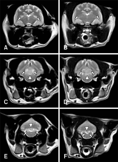
Fig. 1 Transverse T2-weighted MR images (WI) of (A, C, and E) a cat with thiamine deficiency (Case 1). (A) At the level of the thalamus, symmetrical hyperintense signals at the lateral geniculate nuclei (arrows) were noted. (C) Images of the caudal colliculi (arrows) and periaqueductal gray matter (arrowhead) revealed the presence of hyperintense lesions. (E) Bilateral symmetrical hyperintensities were also evident in the medial vestibular nuclei (arrows) and cerebellar nodulus (arrowhead). After 3 weeks of thiamine administration, transverse MR images showed that the lesions had resolved except for ones in the cerebellar nodulus (B, D, and F).
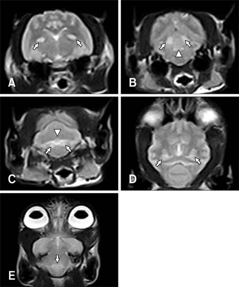
Fig. 2 Transverse (A~C) and dorsal (D and E) T2-WI of a cat with thiamine deficiency (Case 2). (A) Symmetrical hyperintense signals in the lateral geniculate nuclei (arrows) were noted at the level of the thalamus. (B) Images of the caudal colliculi (arrows), periaqueductal gray matter (lesions between the arrows), and facial nuclei (arrowhead) revealed the presence of hyperintense lesions. (C) Bilateral symmetrical hyperintensities were also evident in the medial vestibular nuclei (arrows) and cerebellar nodulus (arrowhead). Bilateral symmetrical hyperintensities were observed in the lateral geniculate body (D; arrows) and cerebral peduncle (E; arrow). After 3 weeks of thiamine administration, clinical signs were nearly resolved. However, additional MRI was not attempted for this cat.
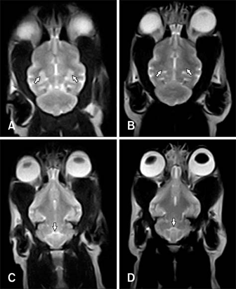
Fig. 3 Dorsal T2-WI of a cat with thiamine deficiency (Case 1). Bilateral, symmetrical hyperintense lesions at the lateral geniculate body (A; arrows) and cerebral peduncle (C; arrow) were observed. Panels B and D correspond to the images shown in panels A and C after 3 weeks of thiamine supplementation. Completely resolved lesions were noted.
Reference
-
1. Davidson MG. Thiamin deficiency in a colony of cats. Vet Rec. 1992; 130:94–97.
Article2. Dreyfus PM, Victor M. Effects of thiamine deficiency on the central nervous system. Am J Clin Nutr. 1961; 9:414–425.
Article3. Garosi LS, Dennis R, Platt SR, Corletto F, de Lahunta A, Jakobs C. Thiamine deficiency in a dog: clinical, clinicopathologic, and magnetic resonance imaging findings. J Vet Intern Med. 2003; 17:719–723.
Article4. Gershoff SN. Nutritional problems of household cats. J Am Vet Med Assoc. 1975; 166:455–458.5. Irle E, Markowitsch HJ. Thiamine deficiency in the cat leads to severe learning deficits and to widespread neuroanatomical damage. Exp Brain Res. 1982; 48:199–208.
Article6. Loew FM, Martin CL, Dunlop RH, Mapletoft RJ, Smith SI. Naturally-occurring and experimental thiamin deficiency in cats receiving commercial cat food. Can Vet J. 1970; 11:109–113.7. Palus V, Penderis J, Jakovljevic S, Cherubini GB. Thiamine deficiency in a cat: resolution of MRI abnormalities following thiamine supplementation. J Feline Med Surg. 2010; 12:807–810.
Article8. Penderis J, McConnell JF, Calvin J. Magnetic resonance imaging features of thiamine deficiency in a cat. Vet Rec. 2007; 160:270–272.
Article9. Singh M, Thompson M, Sullivan N, Child G. Thiamine deficiency in dogs due to the feeding of sulphite preserved meat. Aust Vet J. 2005; 83:412–417.
Article10. Steel RJ. Thiamine deficiency in a cat associated with the preservation of ‘pet meat’ with sulphur dioxide. Aust Vet J. 1997; 75:719–721.
Article
- Full Text Links
-
- Actions
-
Cited
- CITED
-
- Close
- Share
-
- Similar articles
-
- A Case of Wernicke-Korsakoff Syndrome Associated with Hyperemesis Gravidarum
- Wernicke's encephalopathy in a patient with acute alcoholic pancreatitis
- Experiences of Wet Beriberi and Wernicke's Encephalopathy Caused by Thiamine Deficiency in Critically Ill Patients
- Wernicke's Syndrome Induced by Hyperemesis Gravidarum
- Effect of Thiamine Deficiency on Drug Hydroxylation and Level of Cytochrome P-450 in the Rats
