Dorsal Epidural Migration of Lumbar Sequestered Disc Fragment: Report of Three Cases
- Affiliations
-
- 1Department of Radiology, Wonkwang University Hospital, School of Medicine, Wonkwang University, Iksan, Korea. juhngsk@wonkwang.ac.kr
- 2Department of Orthopaedic Surgery, Wonkwang University Hospital, School of Medicine, Wonkwang University, Iksan, Korea.
- KMID: 1709019
- DOI: http://doi.org/10.3348/jksr.2014.71.1.34
Abstract
- Herein we report three cases of dorsal epidural migration of sequestered lumbar disc fragment in male patients older than 60 years. One of the patients underwent a microscopic decompression and discectomy for a herniated lumbar intervertebral disc disease five months ago. The magnetic resonance imaging showed characteristic features of dorsal epidural migration of a sequestered lumbar disc fragment. We suggest that both, older age and previous intervertebral decompression surgery may be the predisposing factors for the dorsal epidural migration of the sequestered lumbar disc fragment.
MeSH Terms
Figure
-

Fig. 1 95 years old male who presented motor weakness of both lower extremities for 2 weeks. A-D. Sagittal (A-C) and axial (D) MR images show an abnormal mass-like lesion located in dorsal aspect to the epidural space at L3/4 level (arrows). The signal intensity of the lesion is low on T2-weighted images (T2WI) (A, D) and intermediate on T1-weighted images (T1WI) (B), similar to signal intensity of adjacent intervertebral disc on both T1WI and T2WI. Following intravenous gadolinium enhancement (C) the lesion shows rim contrast enhancement on fat saturated T1WI. E. The surgical specimen shows fragmented disc material. F. Photomicrograph with hematoxylin and eosin (× 40) stain shows degenerated nucleus pulposus of the disc.
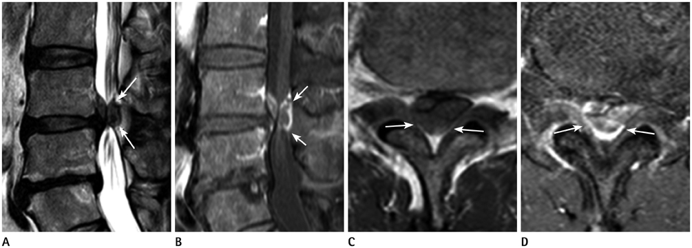
Fig. 2 62 years old male who developed severe pain of low back and both lower extremities for a week after lifting the heavy object. A-D. Sagittal (A-C) and axial (D) MR images show an abnormal mass-like lesion located in dorsal aspect to the epidural space at L4/5 level (arrows). The signal intensity of the lesion is low on T2-weighted images (T2WI) (A, C) and intermediate on T1-weighted images (T1WI) (B), similar to signal intensity of adjacent intervertebral disc on both T1WI and T2WI. Following intravenous gadolinium enhancement (B, D) the lesion shows rim contrast enhancement on fat saturated T1WI.
Reference
-
1. Masaryk TJ, Ross JS, Modic MT, Boumphrey F, Bohlman H, Wilber G. High-resolution MR imaging of sequestered lumbar intervertebral disks. AJR Am J Roentgenol. 1988; 150:1155–1162.2. Bonaroti EA, Welch WC. Posterior epidural migration of an extruded lumbar disc fragment causing cauda equina syndrome. Clinical and magnetic resonance imaging evaluation. Spine (Phila Pa 1976). 1998; 23:378–381.3. Huang TY, Lee KS, Tsai TH, Su YF, Hwang SL. Posterior epidural migration of sequestrated lumbar disc fragments into the bilateral facet joints: case report. Neurosurgery. 2011; 69:E1148–E1151.4. Dösoğlu M, Is M, Gezen F, Ziyal MI. Posterior epidural migration of a lumbar disc fragment causing cauda equina syndrome: case report and review of the relevant literature. Eur Spine J. 2001; 10:348–351.5. Robe P, Martin D, Lenelle J, Stevenaert A. Posterior epidural migration of sequestered lumbar disc fragments. Report of two cases. J Neurosurg. 1999; 90(2):Suppl. 264–266.6. Kuzeyli K, Cakir E, Usul H, Baykal S, Yazar U, Karaarslan G, et al. Posterior epidural migration of lumbar disc fragments: report of three cases. Spine (Phila Pa 1976). 2003; 28:E64–E67.7. Ross JS, Modic MT, Masaryk TJ, Carter J, Marcus RE, Bohlman H. Assessment of extradural degenerative disease with Gd-DTPA-enhanced MR imaging: correlation with surgical and pathologic findings. AJNR Am J Neuroradiol. 1989; 10:1243–1249.8. Yamashita K, Hiroshima K, Kurata A. Gadolinium-DTPA--enhanced magnetic resonance imaging of a sequestered lumbar intervertebral disc and its correlation with pathologic findings. Spine (Phila Pa 1976). 1994; 19:479–482.9. Sandhu FS, Dillon WP. Spinal epidural abscess: evaluation with contrast-enhanced MR imaging. AJNR Am J Neuroradiol. 1991; 12:1087–1093.10. Wiltse LL, Fonseca AS, Amster J, Dimartino P, Ravessoud FA. Relationship of the dura, Hofmann's ligaments, Batson's plexus, and a fibrovascular membrane lying on the posterior surface of the vertebral bodies and attaching to the deep layer of the posterior longitudinal ligament. An anatomical, radiologic, and clinical study. Spine (Phila Pa 1976). 1993; 18:1030–1043.11. Scapinelli R. The meningovertebral ligaments as a barrier to the side-to-side migration of extruded lumbar disc herniations. Acta Orthop Belg. 1992; 58:436–441.
- Full Text Links
-
- Actions
-
Cited
- CITED
-
- Close
- Share
-
- Similar articles
-
- Posterior Epidural Migration of Lumbar Disc Fragment: Three Cases and Review of Literature
- Posterior Epidural Migration of Herniated Lumbar Intervertebral Disc: Two Cases Report
- Posterior Epidural Migration of Lumbar Ruptured Disc: Report of Two Cases
- Posterior Epidural Migration of Thoracic Disc Fragment
- Posterior Migration of Extruded Lumbar Disc Fragments
