T-Condylar Fracture of Distal Humerus in a Child: A Case Report
- Affiliations
-
- 1Department of Orthopaedic Surgery, Samsung Changwon Hospital, Sungkyunkwan University School of Medicine, Changwon, Korea. jeongmin3744@daum.com
- KMID: 1708717
- DOI: http://doi.org/10.12671/jkfs.2014.27.3.232
Abstract
- T-condylar fracture is a type of distal humerus fracture. T-condylar fracture in children is rare, with reported incidence of less than 1% of T-condylar fractures. The mean reported age of T-condylar fracture in children is 11. Cases in children under 5 years-old are extremely rare. Herein, we report on a T-condylar fracture of the distal humerus in a 5-year-old boy. This patient was treated with open reduction and K-wire fixation through the posterolateral approach. The result of treatment was satisfactory; therefore, we report this case.
Keyword
Figure
-

Fig. 1 Initial plain antero-posterior (AP) and lateral view of x-ray. (A) AP view of plain x-ray shows the intercondylar fracture line. (B) Lateral view of plain x-ray shows posterior displacement at the lateral condyle of the left distal humerus.
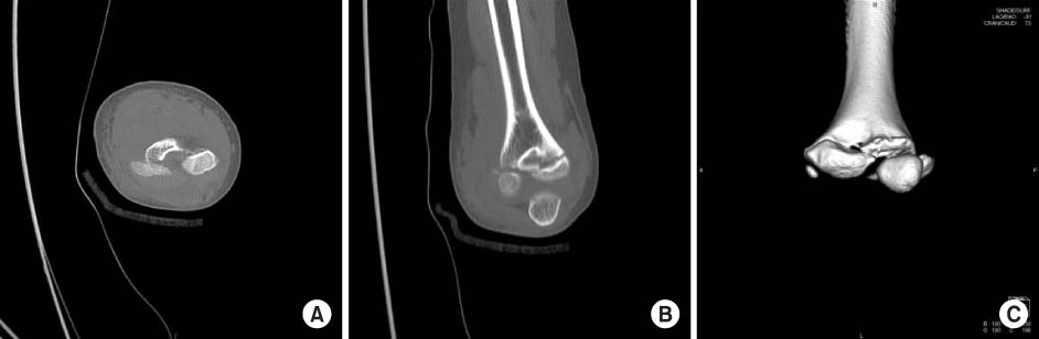
Fig. 2 Pre-operative computed tomography (CT) image of the left elbow. (A) Axial view shows the supracondylar fracture line. (B) Lateral condylar fracture line is definite in the coronal view. (C) T-condylar fracture and posterior displacement of the lateral condyle is remarkable in 3-dimensional CT image.
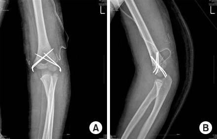
Fig. 3 Plain x-ray after open reduction and Kirschner wire fixation. (A) Antero-posterior view of plain x-ray shows reduced, anatomically. (B) Shaft-condylar angle was recovered on lateral view x-ray.
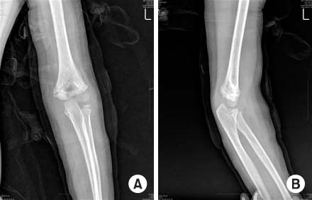
Fig. 4 Five weeks after the operation, Kirschner-wires are removed. (A) Well-bone union is checked in the anteroposterior view of plain x-ray. (B) Posterior displacement is reduced successfully in lateral view of plain x-ray.
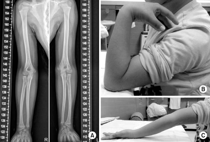
Fig. 5 Two years and seven months after surgery. (A) Valgus or varus deformity is not found in long bone view of plain x-ray. (B) 125 degrees of active elbow flexion was possible. (C) There was no flexion contracture at the elbow.
Reference
-
1. Hasankhani E. T-condylar fracture of distal humerus in 5-year-old child. A report of one case. Iran J Med Sci. 2004; 29:195–197.2. Beghin JL, Bucholz RW, Wenger DR. Intercondylar fractures of the humerus in young children. A report of two cases. J Bone Joint Surg Am. 1982; 64:1083–1087.
Article3. Charles A, Rockwood JR. Fracture in children. 5th ed. Philadelphia: Lippincott-Raven Co;2001. p. 687.4. Mihran O. Pediatric orthopedics. 3rd ed. Philadelphia: WB Saunders Co;2002. p. 2202.5. Papavasiliou VA, Beslikas TA. T-condylar fractures of the distal humeral condyles during childhood: an analysis of six cases. J Pediatr Orthop. 1986; 6:302–305.
Article6. Kanellopoulos AD, Yiannakopoulos CK. Closed reduction and percutaneous stabilization of pediatric T-condylar fractures of the humerus. J Pediatr Orthop. 2004; 24:13–16.
Article7. Re PR, Waters PM, Hresko T. T-condylar fractures of the distal humerus in children and adolescents. J Pediatr Orthop. 1999; 19:313–318.
Article8. Kasser JR, Beaty JH. Rockwood and Wilkins' fractures in children. 5th ed. Philadelphia: Lippincott Williams and Wilkins;2001. p. 577.9. Kantharajanna SB, Goni V, Sudesh P, Gopinathan NR. T-condylar fracture delayed for 10 days in a 5-year-old boy: a case report and review of the literature. Chin J Traumatol. 2013; 16:58–60.
- Full Text Links
-
- Actions
-
Cited
- CITED
-
- Close
- Share
-
- Similar articles
-
- Diagnosis and Treatment of the Lateral Condylar Fracture of Humerus Traversing the Capitulum in Children
- A Comminuted Spiral Fracture with Butterfly Fragment of Distal Humerus by Arm Wrestling: A Case Report
- Radiologic Study of Torsion Type of Supracondylar Fracture of Humerus in Children
- Treatment for Comminuted fractures of Distal End of Humerus by Newly Developed Anatomical Plate: Three case report
- Early Corrective Osteoclasis for Malunited Pediatric Medial Condylar Fracture of the Humerus
