External Ophthalmomyiasis Presenting to an Emergency Department: Corneal Findings as a Sign of Oestrus ovis
- Affiliations
-
- 1Department of Ophthalmology, BirjanUniversity of Medical Science, Birjand, Iran. Yaqubig@yahoo.com
- KMID: 1707287
- DOI: http://doi.org/10.3341/kjo.2013.27.5.341
Abstract
- PURPOSE
This study aims to determine the frequency of opthalmyomyiasis externa and the ocular findings of disease in Southern Khorasan.
METHODS
All patients referred to the emergency department of Valiaser hospital during the year 2011 with external ophthalmomyiasis were enrolled in this study. The diagnosis of external ophthalmomyiasis was made according to clinical findings and the presence of Oestrus ovis larvae.
RESULTS
There were 18 cases of external ophthalmomyiasis in the emergency department of Valiaser hospital in 2011. Most cases had the common signs and symptoms of allergic conjunctivitis, except for three males who were referred with respective complaints of red eye, foreign body sensation, and swelling around the eyelids after contact injury the previous day; corneal infiltration was present in three cases. The visual acuity among the three cases that had peripheral corneal involvement was 20 / 30 in both eyes. The bulbar conjunctiva showed chemosis in all cases and a ropy pattern discharge that was clinically compatible with external ophthalmomyiasis. However, in one case, microscopic slit lamp examination did not show Oestrus ovis larvae.
CONCLUSIONS
The frequency of external ophthalmomyiasis was high in this region. Although external ophthalmomyiasis usually manifests as allergic conjunctivitis, coronary-like corneal infiltration may be considered in the differential diagnosis of external ophthalmomyiasis or toxic insult.
MeSH Terms
-
Adolescent
Adult
Aged
Aged, 80 and over
Animals
Cornea/parasitology/*pathology
Diagnosis, Differential
*Diptera
*Emergency Service, Hospital
Eye Infections, Parasitic/*diagnosis/epidemiology/parasitology
Female
Humans
Incidence
Iran/epidemiology
Larva
Male
Middle Aged
Myiasis/*diagnosis/epidemiology/parasitology
Retrospective Studies
Young Adult
Figure
-
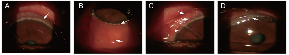
Fig. 1 The first case. Circumb-limbal corneal infilteraton. (A) Superior cornea infiltration. (B) Inferior cornea infiltration and conjunctival chemosis (arrow). (C) Sever conjunctival edema and infiltration (superior arrow) and ropy pattern discharge (inferior arrow). (D) Magnified corneal infilteraton.
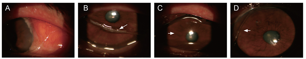
Fig. 2 The second case. (A) The superior peripheral corneal infilteration. (B) Heavy ropy pattern discharge (arrow). (C) Clear cornea centeraly (arrow). (D) Fine ropy pattern discharge (arrow).

Fig. 3 The thired case. (A) Superior corneal infilteration. (B) Inferior corneal infilteration. (C) Lived Oestrus ovis in conjunctiva. (D) Dead Oestrus ovis in lid (arrow).
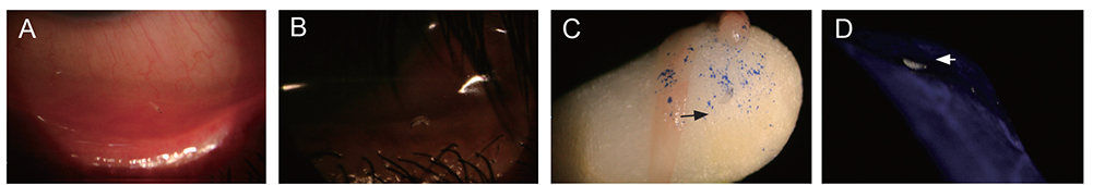
Fig. 4 (A,B) The whole larva with translucent segmented body and large dark oral hooks connected to a cephalopharyngeal skeleton (arrow). (C,D) Magnified view will showing the anterior part of the dead maggot with a pair of dark oral hooks (arrow).
Reference
-
1. Gregory AR, Schatz S, Laubach H. Ophthalmomyiasis caused by the sheep bot fly Oestrus ovis in northern Iraq. Optom Vis Sci. 2004; 81:586–590.2. Gupta VP, Baveja UK. Ophthalmomyiasis externa caused by the sheep nasal botfly Oestrus ovis. Indian J Ophthalmol. 1988; 36:41–42.3. Sreejith RS, Reddy AK, Ganeshpuri SS, Garg P. Oestrus ovis ophthalmomyiasis with keratitis. Indian J Med Microbiol. 2010; 28:399–402.4. Misra S, Misra N, Reddy B. External ophthalmomyiasis by Oestrus ovis: an unknown endemic eye disease in rural parts of central India. Trop Doct. 2008; 38:120–122.5. Abdellatif MZ, Elmazar HM, Essa AB. Oestrus ovis as a cause of red eye in Aljabal Algharbi, Libya. Middle East Afr J Ophthalmol. 2011; 18:305–308.6. Jenzeri S, Ammari W, Attia S, et al. External ophthalmomyiasis manifesting with keratouveitis. Int Ophthalmol. 2009; 29:533–535.7. Thakur K, Singh G, Chauhan S, Sood A. Vidi, vini, vinci: external ophthalmomyiasis infection that occurred, and was diagnosed and treated in a single day: a rare case report. Oman J Ophthalmol. 2009; 2:130–132.8. Kanski JJ. Disorder of conjunctiva. In : Kanski JJ, editor. Clinical ophthalmology. 4th ed. Oxford: Butterworth-Heinemann;1999. p. 59–93.9. Masoodi M, Hosseini K. External ophthalmomyiasis caused by sheep botfly (Oestrus ovis) larva: a report of 8 cases. Arch Iranian Med. 2004; 7:136–139.10. Sharifipour F, Feghhi M. Anterior ophthalmomyiasis interna: an ophthalmic emergency. Arch Ophthalmol. 2008; 126:1466–1467.11. Nigwekar S. Ophthalmomyiasis externa: a case report. Pravara Med Rev. 2009; 4:28–30.
- Full Text Links
-
- Actions
-
Cited
- CITED
-
- Close
- Share
-
- Similar articles
-
- External Ophthalmomyiasis Caused by Oestrus ovis: A Rare Case Report from India
- A Case of Recurrent External Ophthalmomyiasis Caused by Lucilia sericata Meigen in a Healthy Patient
- Corneal Foreign Body Removal by Emergency Physicians
- Protection against virulent Brucella spp. by gamma-irradiated B. ovis in BALB/c mice model
- Comparison between two progesterone sources and two oestradiol formulations in a Heatsynch protocol for postpartum cycling dairy cows in pasture
