Korean J Ophthalmol.
2013 Oct;27(5):311-315. 10.3341/kjo.2013.27.5.311.
Ptosis Repair Using Preserved Fascia Lata with the Modified Direct Tarsal Fixation Technique
- Affiliations
-
- 1Department of Ophthalmology, Dong-A University Medical Center, Busan, Korea. hbahn@dau.ac.kr
- KMID: 1707283
- DOI: http://doi.org/10.3341/kjo.2013.27.5.311
Abstract
- PURPOSE
To evaluate the clinical outcome of frontalis sling operation using preserved fascia lata with modified direct tarsal fixation in congenital ptosis patients.
METHODS
Forty-seven congenital ptosis patients (60 eyes) who underwent a frontalis sling operation using preserved fascia lata with modified direct tarsal fixation method between March 2001 and December 2008 with a mean follow-up time of 52 months (range, 26 to 122 months) were included in this study. The medical records were reviewed retrospectively.
RESULTS
A retrospective chart review was conducted in patients who were diagnosed with congenital ptosis and underwent frontalis suspension surgery using preserved fascia lata with modified direct tarsal fixation from 2001 through 2008 at Dong-A University Hospital. The patients were 34 males and 14 females. The age of the patients ranged from 1 to 18 years with an average age of 4.51 years. At a mean follow-up of 60 months, good final results were achieved in 46 eyes (76.6%), fair in 8 eyes (13.3%), and poor in 6 eyes (10%). The poor results consisted of undercorrection of 1 eye and recurrence in 5 eyes. The accumulative survival rate was 87.2%, with all recurrences occurring within 12 months postoperatively.
CONCLUSIONS
Frontalis sling operation by preserved fascia lata with modified direct tarsal fixation appears to be an effective treatment for severe congenital ptosis, showing good long term results.
Keyword
MeSH Terms
Figure
-
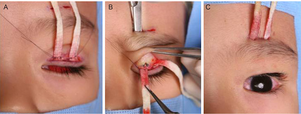
Fig. 1 (A) The ends of the two pieces of fascia lata were anchored to the upper tarsus with 6-0 nylon suture. (B) A Kelly was inserted into the suprabrow incision, grabbing the free end of the sutured fascia lata. (C) The ends of fascia lata were pulled through the sides of the suprabow incisions.
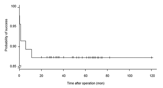
Fig. 2 The cumulative survival curve. The cumulative survival rate was 87.2%. Recurrence occurred after a mean of 3.7 ± 4.6 months.
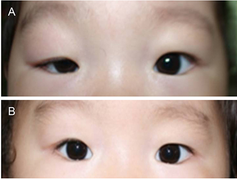
Fig. 3 Pictures of one patient before (A) and after (B) the surgery. The degree of ptosis was moderate and surgical outcome was good.
Reference
-
1. Beard C. Ptosis. 3rd ed. St. Louis: Mosby;1981. p. 169–173.2. Mustarde JC. Repair and reconstruction in the orbital region. 2nd ed. Edinburgh: Churchill Livingstone;1980. p. 325–328.3. Fox SA. Congenital ptosis II. Frontalis sling. J Paediatr Ophthalmol. 1966; 3:25.4. Song R, Song Y. Treatment of blepharoptosis: direct transplantation of the frontalis muscle to the upper eyelid. Clin Plast Surg. 1982; 9:45–48.5. Crawford JS. Recent trends in ptosis surgery. Ann Ophthalmol. 1975; 7:1263–1267.6. Fox SA. A new frontalis skin sling for ptosis. Am J Ophthalmol. 1968; 65:359–362.7. Crawford JS. Repair of ptosis using frontalis muscle and fascia lata. Trans Am Acad Ophthalmol Otolaryngol. 1956; 60:672–678.8. Goldberger S, Conn H, Lemor M. Double rhomboid silicone rod frontalis suspension. Ophthal Plast Reconstr Surg. 1991; 7:48–53.9. Seider N, Beiran I, Kaltreider SA. One medial triangular Tutoplast sling as a frontalis suspension for adult myogenic blepharoptosis. Acta Ophthalmol Scand. 2006; 84:121–123.10. Mauriello JA Jr, Abdelsalam A. Effectiveness of homologous cadaveric fascia lata and role of suture fixation to tarsus in frontalis suspension. Ophthal Plast Reconstr Surg. 1998; 14:99–104.11. Spoor TC, Kwitko GM. Blepharoptosis repair by fascia lata suspension with direct tarsal and frontalis fixation. Am J Ophthalmol. 1990; 109:314–317.12. Crawford JS. Repair of ptosis using frontalis muscle and fascia lata: a 20-year review. Ophthalmic Surg. 1977; 8:31–40.13. Crawford JS. Frontalis sling operation. J Pediatr Ophthalmol Strabismus. 1982; 19:253–255.14. Wagner RS, Mauriello JA Jr, Nelson LB, et al. Treatment of congenital ptosis with frontalis suspension: a comparison of suspensory materials. Ophthalmology. 1984; 91:245–248.15. Wasserman BN, Sprunger DT, Helveston EM. Comparison of materials used in frontalis suspension. Arch Ophthalmol. 2001; 119:687–691.16. Kemp EG, James CR, Collin JR. Brow suspension in the management of ptosis: an analysis of over 100 cases. Trans Ophthalmol Soc U K. 1986; 105(Pt 1):84–87.17. El-Toukhy E, Salaem M, El-Shewy T, et al. Mersilene mesh sling as an alternative to autogenous fascia lata in the management of ptosis. Eye (Lond). 2001; 15(Pt 2):178–182.18. Leibovitch I, Leibovitch L, Dray JP. Long-term results of frontalis suspension using autogenous fascia lata for congenital ptosis in children under 3 years of age. Am J Ophthalmol. 2003; 136:866–871.19. Beyer CK, Albert DM. The use and fate of fascia lata and sclera in ophthalmic plastic and reconstructive surgery. Ophthalmology. 1981; 88:869–886.20. Wilson ME, Johnson RW. Congenital ptosis: long-term results of treatment using lyophilized fascia lata for frontalis suspensions. Ophthalmology. 1991; 98:1234–1237.21. Broughton WL, Matthews JG 2nd, Harris DJ Jr. Congenital ptosis: results of treatment using lyophilized fascia lata for frontalis suspensions. Ophthalmology. 1982; 89:1261–1266.
- Full Text Links
-
- Actions
-
Cited
- CITED
-
- Close
- Share
-
- Similar articles
-
- Congenital Ptosis Repair by Preserved Fascia Lata with Direct Tarsal and Frontalis Fixation
- Frontalis Sling Operation by Fixation of Preserved Fascia Lata to Tarsus in Congenital Blepharoptosis Patients
- The Frontalis Sling Operation Using Preserved Fascia Lata: Modified Crawford Technique
- Frontalis Suspension Using Preserved Fascia Lata for Severe Congenital Ptosis Under 2 years of Age
- The effect of Frontalis suspension Ptosis repair using Fascia lata in congenital unilateral ptosis
