Evaluation of changes in left ventricular myocardial function observed in canine myocardial dysfunction model using a two-dimensional tissue tracking technique
- Affiliations
-
- 1Department of Veterinary Surgery, Faculty of Veterinary Medicine, Tokyo University of Agriculture and Technology, Tokyo 183-8509, Japan. ryo@vet.ne.jp
- 2Department of Veterinary Internal Medicine, Faculty of Veterinary Medicine, Tokyo University of Agriculture and Technology, Tokyo 183-8509, Japan.
- KMID: 1705567
- DOI: http://doi.org/10.4142/jvs.2013.14.3.355
Abstract
- This study was conducted to assess the ability of two-dimensional tissue tracking (2DTT) to evaluate changes in left ventricular (LV) myocardial function associated with sustained high electrical pacing. Pacemakers were implanted at the right ventricular (RV) apex of five female Beagles, and sustained high electrical pacing of 250 beats per minute (bpm) was performed for three consecutive weeks. Conventional echocardiography and 2DTT were performed at baseline, and at every week for three weeks with pacing. The baseline parameters were then compared to those of weeks 1, 2, and 3. Three weeks of pacing resulted in significant reduction of radial and circumferential global strains (p < 0.001). Regional analysis revealed reduction of segmental strains in both radial and circumferential directions, as well as increased dyssynchrony after three weeks of pacing in the radial direction (p = 0.0007). The results of this study revealed the ability of 2DTT to measure radial and circumferential strains in dogs with sustained high-electrical pacing, and allowed assessment of global and regional myocardial function and the degree of dyssynchrony.
MeSH Terms
Figure
-

Fig. 1 Examples of radial and circumferential strain profiles obtained from the right parasternal short axis view at the level of the papillary muscle using two-dimensional tissue tracking. Radial strain represents thickening and thinning motion of the myocardium in the radial direction, and circumferential strain represents myocardial motion along the circular perimeter. In the systole, the myocardial deformation increases in the radial direction, thus the radial strain becomes positive, and the myocardial deformation decreases in the circumferential direction, causing the circumferential direction to becomes negative. Radial (A) and circumferential (B) strains at baseline, and radial (C) and circumferential (D) strains at week 3 of sustained high electrical pacing.
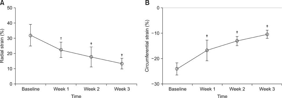
Fig. 2 Mean ± standard deviation values of global radial (A) and circumferential (B) strains plotted against time. Compared with baseline, †p < 0.01, ‡p <0.001 (ANOVA-Dunnet's post test).
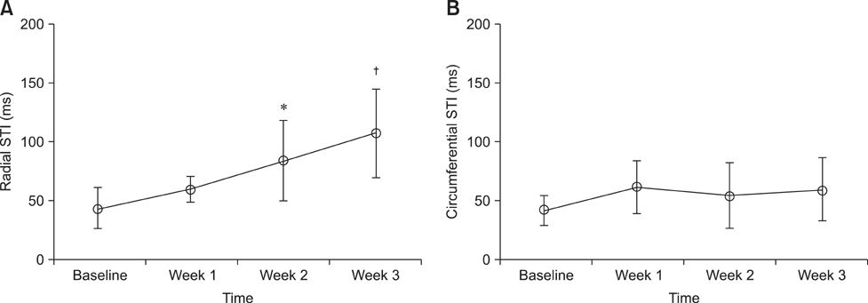
Fig. 3 Mean ± standard deviation values of radial (A) and circumferential (B) synchrony time index, which is the indicator used to assess the degree of dyssynchrony. Significant differences determined by Dunnett's multiple comparison tests are represented when relevant. Compared with baseline, *p < 0.05, †p < 0.001.
Reference
-
1. Amundsen BH, Helle-Valle T, Edvardsen T, Torp H, Crosby J, Lyseggen E, Stoylen A, Ihlen H, Lima JA, Smiseth OA, Slordahl SA. Noninvasive myocardial strain measurement by speckle tracking echocardiography: validation against sonomicrometry and tagged magnetic resonance imaging. J Am Coll Cardiol. 2006; 47:789–793.
Article2. Arita T, Sorescu GP, Schuler BT, Schmarkey LS, Merlino JD, Vinten-Johansen J, Leon AR, Martin RP, Sorescu D. Speckle-tracking strain echocardiography for detecting cardiac dyssynchrony in a canine model of dyssynchrony and heart failure. Am J Physiol Heart Circ Physiol. 2007; 293:H735–H742.
Article3. Artis NJ, Oxborough DL, Williams G, Pepper CB, Tan LB. Two-dimensional strain imaging: a new echocardiographic advance with research and clinical applications. Int J Cardiol. 2008; 123:240–248.
Article4. Becker M, Lenzen A, Ocklenburg C, Stempel K, Kühl H, Neizel M, Katoh M, Kramann R, Wildberger J, Kelm M, Hoffmann R. Myocardial deformation imaging based on ultrasonic pixel tracking to identify reversible myocardial dysfunction. J Am Coll Cardiol. 2008; 51:1473–1481.
Article5. Boon JA. Veterinary Echocardiography. 2nd ed. Chichester: John Wiley & Sons;2011. p. 101–112.6. Chetboul V. Advanced techniques in echocardiography in small animals. Vet Clin North Am Small Anim Pract. 2010; 40:529–543.
Article7. Chetboul V, Sampedrano CC, Gouni V, Nicolle AP, Pouchelon JL, Tissier R. Ultrasonographic assessment of regional radial and longitudinal systolic function in healthy awake dogs. J Vet Intern Med. 2006; 20:885–893.
Article8. Chetboul V, Serres F, Gouni V, Tissier R, Pouchelon JL. Radial strain and strain rate by two-dimensional speckle tracking echocardiography and the tissue velocity based technique in the dog. J Vet Cardiol. 2007; 9:69–81.
Article9. Culwell NM, Bonagura JD, Schober KE. Comparison of echocardiographic indices of myocardial strain with invasive measurements of left ventricular systolic function in anesthetized healthy dogs. Am J Vet Res. 2011; 72:650–660.
Article10. Dandel M, Hetzer R. Echocardiographic strain and strain rate imaging--clinical applications. Int J Cardiol. 2009; 132:11–24.11. Edvardsen T, Helle-Valle T, Smiseth OA. Systolic dysfunction in heart failure with normal ejection fraction: speckle-tracking echocardiography. Prog Cardiovasc Dis. 2006; 49:207–214.
Article12. Friedberg MK, Slorach C. Relation between left ventricular regional radial function and radial wall motion abnormalities using two-dimensional speckle tracking in children with idiopathic dilated cardiomyopathy. Am J Cardiol. 2008; 102:335–339.
Article13. Fuentes VL. Echocardiography and Doppler ultrasound. In : Tilley L, Smith F, Oyama M, Sleeper M, editors. Manual of Canine and Feline Cardiology. 4th ed. Missouri: Saunders;2008. p. 78–98.14. Geyer H, Caracciolo G, Abe H, Wilansky S, Carerj S, Gentile F, Nesser HJ, Khandheria B, Narula J, Sengupta PP. Assessment of myocardial mechanics using speckle tracking echocardiography: fundamentals and clinical applications. J Am Soc Echocardiogr. 2010; 23:351–369.
Article15. Labombarda F, Blanc J, Pellissier A, Stos B, Gaillard C, Bajolle F, Maltret A, Sidi D, Bonnet D, Boudjemline Y. Health-e-Child project: mechanical dyssynchrony in children with dilated cardiomyopathy. J Am Soc Echocardiogr. 2009; 22:1289–1295.
Article16. Lindqvist P, Borgström E, Gustafsson U, Mörner S, Henein MY. Asynchronous normal regional left ventricular function assessed by speckle tracking echocardiography: appearances can be deceptive. Int J Cardiol. 2009; 134:195–200.
Article17. Mondillo S, Galderisi M, Mele D, Cameli M, Lomoriello VS, Zacà V, Ballo P, D’Andrea A, Muraru D, Losi M, Agricola E, D'Errico A, Buralli S, Sciomer S, Nistri S, Badano L. Speckle-tracking echocardiography: a new technique for assessing myocardial function. J Ultrasound Med. 2011; 30:71–83.18. Nesbitt GC, Mankad S, Oh JK. Strain imaging in echocardiography: methods and clinical applications. Int J Cardiovasc Imaging. 2009; 25:Suppl 1. 9–22.
Article19. Nesser HJ, Winter S. Speckle tracking in the evaluation of left ventricular dyssynchrony. Echocardiography. 2009; 26:324–336.
Article20. Perk G, Tunick PA, Kronzon I. Non-Doppler two-dimensional strain imaging by echocardiography - from technical considerations to clinical applications. J Am Soc Echocardiogr. 2007; 20:234–243.
Article21. Schwarzwald CC, Schober KE, Berli ASJ, Bonagura JD. Left ventricular radial and circumferential wall motion analysis in horses using strain, strain rate, and displacement by 2D speckle tracking. J Vet Intern Med. 2009; 23:890–900.
Article22. Shinbane JS, Wood MA, Jensen DN, Ellenbogen KA, Fitzpatrick AP, Scheinman MM. Tachycardia-induced cardiomyopathy: a review of animal models and clinical studies. J Am Coll Cardiol. 1997; 29:709–715.
Article23. Takano H, Fujii Y, Ishikawa R, Aoki T, Wakao Y. Comparison of left ventricular contraction profiles among small, medium, and large dogs by use of two-dimensional speckle-tracking echocardiography. Am J Vet Res. 2010; 71:421–427.
Article24. Tomaske M, Breithardt OA, Balmer C, Bauersfeld U. Successful cardiac resynchronization with single-site left ventricular pacing in children. Int J Cardiol. 2009; 136:136–143.
Article25. Tops LF, Suffoletto MS, Bleeker GB, Boersma E, van der Wall EE, Gorcsan J 3rd, Schalij MJ, Bax JJ. Speckle-tracking radial strain reveals left ventricular dyssynchrony in patients with permanent right ventricular pacing. J Am Coll Cardiol. 2007; 50:1180–1188.
Article26. Wang J, Khoury DS, Yue Y, Torre-Amione G, Nagueh SF. Preserved left ventricular twist and circumferential deformation, but depressed longitudinal and radial deformation in patients with diastolic heart failure. Eur Heart J. 2008; 29:1283–1289.
Article27. Wess G, Keller LJM, Klausnitzer M, Killich M, Hartmann K. Comparison of longitudinal myocardial tissue velocity, strain, and strain rate measured by two-dimensional speckle tracking and by color tissue Doppler imaging in healthy dogs. J Vet Cardiol. 2011; 13:31–43.
Article28. Wilson JR, Douglas P, Hickey WF, Lanoce V, Ferraro N, Muhammad A, Reichek N. Experimental congestive heart failure produced by rapid ventricular pacing in the dog: cardiac effects. Circulation. 1987; 75:857–867.
Article29. Wu W, Wang H, Tang Y, Yuan W, Wang H, Jiang Y. Application of quantitative tissue velocity imaging to evaluate left ventricular early diastolic dysfunction in dogs with heart failure due to rapid ventricular pacing. J Am Soc Echocardiogr. 2008; 21:1269–1276.
Article30. Yoshikawa H, Suzuki M, Tezuka N, Otsuka T, Sugi K. Differences in left ventricular dyssynchrony between high septal pacing and apical pacing in patients with normal left ventricular systolic function. J Cardiol. 2010; 56:44–50.
Article31. Zupan I, Rakovec P, Budihna N, Brecelj A, Koželj M. Tachycardia induced cardiomyopathy in dogs; relation between chronic supraventricular and chronic ventricular tachycardia. Int J Cardiol. 1996; 56:75–81.
Article
- Full Text Links
-
- Actions
-
Cited
- CITED
-
- Close
- Share
-
- Similar articles
-
- The Usefulness of Doppler Tissue Image in Evaluation of Left Ventricular Systolic and Diastolic Dysfunction
- Assessment of Myocardial Viability Using PET
- Cardiac Strain Analysis Using Cine Magnetic Resonance Imaging and Computed Tomography
- Current Status of 3-Dimensional Speckle Tracking Echocardiography: A Review from Our Experiences
- Principles and Clinical Applications of Feature-Tracking Cardiac Magnetic Resonance Imaging: A Literature Review
