Effects of intertrochanteric varus osteotomy on Norberg angle and percent coverage of the femoral head in displastic dogs
- Affiliations
-
- 1Department of Veterinary Clinical Sciences, University of Bologna, 40064 Ozzano Emilia, Italy. stefania.pinna@unibo.it
- KMID: 1705520
- DOI: http://doi.org/10.4142/jvs.2013.14.2.185
Abstract
- This study was conducted to assess the effects of femoral varus osteotomy on joint congruency in dogs affected by early stage hip dysplasia. Preoperative planning to move the femoral head within the acetabulum was carried out. Varisation of the femoral inclination angle (fIA) was achieved by Intertrochanteric Osteotomy (ITO). Norberg angle (NA), percent coverage (PC) of the femoral head by the acetabulum and fIA was measured from preoperative, immediate postoperative and first and second recheck radiographs of seven dogs that underwent an ITO (joint n = 9). There was significant (p < 0.05) improvement of both NA and PC in all patients as indicated by a change in the mean +/- standard deviation of 78.9degrees +/- 7.5 and 36.9% +/- 5.2 to 92.2degrees +/- 6.7 and 50.6% +/- 8.3, respectively. No significant difference (p < 0.05) was observed between the values of the planned femoral inclination angle (pfIA) of the femur and the effective femoral inclination angle (efIA) obtained after surgery (115.9degrees +/- 2.5 and 111.3degrees +/- 6.4, respectively). These findings could encourage the use of ITO in veterinary practice and indicate that intertrochanteric varus osteotomy should be re-considered for the treatment of early stage hip dysplasia in dogs with radiological signs of joint incongruency.
MeSH Terms
Figure
-
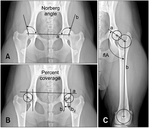
Fig. 1 X-ray images of a 14 month old female Border Collie (21 kg) with B1 hip dysplasia. (A) Norberg angle is measured on the radiograph and indicates the amount of joint laxity. (a) Line between the center points on each of the two femoral heads; (b) line between the center of the femoral head and the craniolateral aspect of the acetabular rim. (B) The percent coverage (PC) of the femoral head by the acetabulum is measured as described by Belkoff. PC is an indication of the support provided by the acetabulum to oppose the force transmitted from the femur. (a) Line between the two craniolateral aspects of the acetabular rims; (b1) line touching the medial surface of the femoral head; (b2) line touching the cranial acetabular rim; (c) femoral head diameters. The overlap distances, b1 and b2, are divided by the femoral head diameter to yield the percentage of the femoral head within the acetabulum. (C) Femoral inclination angle (fIA) determined by the Symax method. (a) Line between the center of the head and the center of the circle drawn at the proximal extremities of the femur; (b) line between the center of the circles drawn at the proximal and distal extremities of the femur.

Fig. 2 X-ray images of an 11 month old female Maremmano-Abruzzese Sheppard (44 kg) with hip joint incongruence. (A) pfIA: planned femoral inclination angle. (B) fIA: femoral inclination angle. CA1: planned correction angle. (C) CA2: shifted correction angle and base of the bony wedge to be removed (arrow).

Fig. 3 X-ray images of an 11 month old Maremmano-Abruzzese Sheppard (44 kg) taken prior to and after ITO in a standard ventrodorsal projection with the femurs parallel and intrarotated. An illustration of changes between the two point times. (A and A1) fIA and efIA, (B and B1) Norberg angle, (C and C1) percentage of coverage, pre and immediately postoperative, respectively.

Fig. 4 X-ray images of a 10 month old male Border Collie (24 kg) taken at each evaluation point in standard ventrodorsal projection with the femurs parallel and intrarotated. The perimeter of the femoral head is in the white circle, while the acetabulum is in the black circle. (A) Preoperative radiograph. Joint is not congruent, the two circles are not within one another and the centers are not coincident. (B) Immediately postoperative radiograph. The circles are congruent and the centers are almost coincident. The osteotomy line is visible. (C) One month postoperative. The position of the femoral head within the acetabulum is preserved; the osteotomy line is still visible. (D) Six months postoperative. The two circles are within each other, indicating joint congruency. The osteotomy line is not visible. The implant is in the right site.
Reference
-
1. Arnoczky SP, Torzilli PA. Biomechanical analysis of forces acting about the canine hip. Am J Vet Res. 1981; 42:1581–1585.2. Banfield CM, Bartels JE, Hudson JA, Wright JC, Hathcock JT, Montgomery RD. A retrospective study of canine hip dysplasia in 116 military working dogs. Part I: Angle measurements and orthopedic foundation for animals (OFA) grading. J Am Anim Hosp Assoc. 1996; 32:413–422.
Article3. Belkoff SM, Padgett G, Soutas-Little RW. Development of a device to measure canine coxofemoral joint laxity. Vet Comp Orthop Traumatol. 1989; 2:31–36.
Article4. Bernardé A. Juvenile pubic symphysiodesis and juvenile pubic symphysiodesis associated with pectineus myotomy: short-term outcome in 56 dysplastic puppies. Vet Surg. 2010; 39:158–164.
Article5. Borostyankoi F, Rooks RL, Kobluk CL, Reed AL, Littledike ET. Results of single-session bilateral triple pelvic osteotomy with an eight-hole iliac bone plate in dogs: 95 cases (1996-1999). J Am Vet Med Assoc. 2003; 222:54–59.
Article6. Braden TD, Prieur WD. Three-plane intertrochanteric osteotomy for treatment of early stage hip dysplasia. Vet Clin North Am Small Anim Pract. 1992; 22:623–643.
Article7. Braden TD, Prieur WD, Kaneene JB. Clinical evaluation of intertrochanteric osteotomy for treatment of dogs with early-stage hip dysplasia: 37 cases (1980-1987). J Am Vet Med Assoc. 1990; 196:337–341.8. Comhaire FH, Criel ACC, Dassy CAA, Guévar PGJ, Snaps FR. Precision, reproducibility, and clinical usefulness of measuring the Norberg angle by means of computerized image analysis. Am J Vet Res. 2009; 70:228–235.
Article9. Dejardin LM, Perry RL, Arnoczky SP. The effect of triple pelvic osteotomy on the articular contact area of the hip joint in dysplastic dogs: an in vitro experimental study. Vet Surg. 1998; 27:194–202.
Article10. Dejardin LM, Perry RL, Arnoczky SP, Torzilli PA. The effect of triple pelvic osteotomy on hip force in dysplastic dogs: a theoretical analysis. Vet Surg. 1996; 25:114–120.
Article11. Evers P, Kramek BA, Wallace LJ, Johnston GR, King V. Clinical and radiographic evaluation of intertrochanteric osteotomy in dogs: a retrospective study of 18 dogs. Vet Surg. 1997; 26:217–222.
Article12. Hauptman J. Interobserver variation in the measurement of the femoral angle of inclination. Vet Surg. 1983; 12:189–191.
Article13. Hauptman J, Prieur WD, Butler HC, Guffy MM. The angle of inclination of the canine femoral head and neck. Vet Surg. 1979; 8:74–77.
Article14. Hauptman J, Cardinet GH 3rd, Morgan JP, Guffy MM, Wallace LJ. Angles of inclination and anteversion in hip dysplasia in the dog. Am J Vet Res. 1985; 46:2033–2036.15. Johnson AL, Smith CV, Pijanowsky GJ, Hungerford LL. Triple pelvic osteotomy: effect on limb function and progression of degenerative joint disease. J Am Anim Hosp Assoc. 1998; 34:260–264.
Article16. Manley PA, Adams WM, Danielson KC, Dueland RT, Linn KA. Long-term outcome of juvenile pubic symphysiodesis and triple pelvic osteotomy in dogs with hip dysplasia. J Am Vet Med Assoc. 2007; 230:206–210.
Article17. Minagawa H, Aiga A, Endo H, Mitani S, Tetsunaga T, Ozaki T. Radiological and clinical results of rotational acetabular osteotomy combined with femoral intertrochanteric osteotomy for avascular necrosis following treatment for developmental dysplasia of the hip. Acta Med Okayama. 2009; 63:169–175.18. Olsson SE. Roentgen examination of the hip joints of German Shepherd dogs. Adv Small Anim Pract. 1961; 3:117–120.19. Poss R. The role of osteotomy in the treatment of osteoarthritis of the hip. J Bone Joint Surg Am. 1984; 66:144–151.
Article20. Prieur WD. Coxarthrosis in dog part I: Normal and abnormal biomechanics of the hip joint. Vet Surg. 1980; 9:145–149.
Article21. Prieur WD. Intertrochanteric osteotomy in the dog: theoretical consideration and operative technique. J Small Anim Pract. 1987; 28:3–20.
Article22. Rasmussen LM, Kramek AB, Lipowitz AJ. Preoperative variables affective long-term outcome of triple pelvic osteotomy for treatment of naturally developing hip dysplasia in dogs. J Am Vet Med Assoc. 1998; 213:80–85.23. Rumph PF, Hathcock JT. A symmetric axis-based method for measuring the projected femoral angle of inclination in dogs. Vet Surg. 1990; 19:328–333.
Article24. Sarierler M. Comparison of femoral inclination angle measurements in dysplastic and nondysplastic dogs of different breeds. Acta Vet Hung. 2004; 52:245–252.
Article25. Schrader SC. Triple osteotomy of the pelvis as a treatment for canine hip dysplasia. J Am Vet Med Assoc. 1981; 178:39–44.26. Schulz KS, Dejardin LM. Surgical treatment of canine hip dysplasia. In : Slatter DH, editor. Textbook of small animal surgery. 3rd ed. Philadelphia: Saunders;2003. p. 2029–2058.27. Slocum B, Slocum TD. Pelvic osteotomy for axial rotation of acetabular segment in dogs with hip dysplasia. Vet Clin North Am Small Anim Pract. 1992; 22:645–682.
Article28. Slocum B, Slocum TD. The Hip. In : Bojrab MJ, Slocum B, Ellison GW, editors. Current technique in small animal surgery. 4th ed. Baltimore: Lippincott Williams & Wilkins;1998. p. 1127–1165.29. Swainson SW, Conzemius MG, Riedesel EA, Smith GK, Riley CB. Effect of pubic symphysiodesis on pelvic development in the skeletally immature greyhound. Vet Surg. 2000; 29:178–190.
Article30. Todhunter RJ, Lust G. Hip Dysplasia: pathogenesis. In : Slatter DH, editor. Textbook of small animal surgery. 3rd ed. Philadelphia: Saunders;2003. p. 2009–2018.31. Tomlinson JL, Cook JL. Effects of degree of acetabular rotation after triple pelvic osteotomy on the position of the femoral head in relationship to the acetabulum. Vet Surg. 2002; 31:398–403.
Article32. Tomlinson JL, Johnson JC. Quantification of measurement of femoral head coverage and Norberg angle within and among four breeds of dog. Am J Vet Res. 2000; 61:1492–1500.
Article33. Vezzoni A, Boiocchi S, Vezzoni L, Vanelli AB, Bronzo V. Double pelvic osteotomy for the treatment of hip dysplasia in young dogs. Vet Comp Orthop Traumatol. 2010; 23:444–452.
Article34. Walker TL, Prieur WD. Intertrochanteric femoral osteotomy. Semin Vet Med Surg (Small Anim). 1987; 2:117–130.35. Zweifel J, Hönle W, Schuh A. Long-term results of intertrochanteric varus osteotomy for dysplastic osteoarthritis of the hip. Int Orthop. 2011; 35:9–12.
Article
- Full Text Links
-
- Actions
-
Cited
- CITED
-
- Close
- Share
-
- Similar articles
-
- Femoral Varus Osteotomy Versus Salter Innominate Osteotomy in the Treatment of Legg - Calve - Perthes Disease
- Femoral Varus Derotation Osteotomy in the Treatment of Acetabular Cysplasia in Deveolpinetal Dysplasia of the Hip
- Pelvic Osteotomy in Adults
- Femoral Osteotomy for Residual Subluxation of Hip after Reduction of Congenital Dislocation
- Comparative Study on the Results of Femoral Osteotomy and Innominate Osteotomy in LCPD
