Kimura Disease Involving a Caruncle
- Affiliations
-
- 1Department of Ophthalmology, Chonbuk National University Hospital, Chonbuk National University Medical School, Jeonju, Korea. you2ic@paran.com
- 2Department of Pathology, Chonbuk National University Hospital, Chonbuk National University Medical School, Jeonju, Korea.
- KMID: 1503808
- DOI: http://doi.org/10.3341/kjo.2013.27.2.137
Abstract
- A 35-year-old woman presented with history of a painless, slow-growing nodule in a left eye caruncle over the last 2 months. The visual acuity was 20 / 20 and the ocular movements were in the normal range. The venereal disease research laboratory test, erythrocyte sedimentation rate, serum angiotensin converting enzyme level, and chest radiograph were all normal. An excisional biopsy was done to confirm the diagnosis. A 1.3 x 0.5 x 0.3 cm sized nodule was extracted and sent for histopathologic examination. Hematoxylin-eosin staining showed follicular hyperplasia with perifollicular fibrosis, an eosinophil infiltrate, and proliferation of capillary vessels. The capillaries were lined by normal-appearing, flat, spindle-shaped endothelial cells. On the basis of these histopathologic findings, the diagnosis of Kimura disease in a caruncle was established. This is the first report describing Kimura disease localized to a caruncle. Kimura disease should be suspected and included in the differential diagnosis of a caruncular mass lesion.
Keyword
MeSH Terms
Figure
-

Fig. 1 A slit lamp biomicroscopic finding on the patient's initial visit revealed a conjunctival nodule in the left medial canthal area.
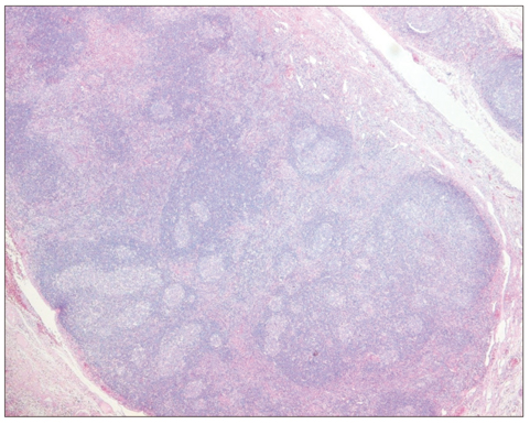
Fig. 2 Histological findings of mass lesion (hematoxylin-eosin stain, ×100). Chronic inflammation with multiple follicular lymphoid hyperplasia with nodal germinal center.
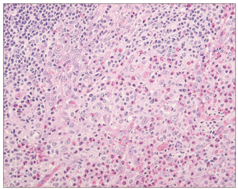
Fig. 3 Numerous infiltrated eosinophils with fibrous septa (hematoxylin-eosin stain, ×400).
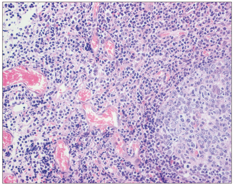
Fig. 4 Multiple capillary proliferation with normal flat, spindle-shaped vascular endothelium (hematoxylin-eosin stain, ×400).

Fig. 5 Immunohistochemical staining for CD20 and CD3 in Kimura disease (×200). The germinal center B-lymphocytes of follicles is positive for CD20 (A) and CD3 stained in the surrounding mantle zone of T-lymphocytes (B).
Reference
-
1. Kimura T, Yoshimura S, Ishikawa E. On the unusual granulation combined with hyperplastic changes of lymphatic tissue. Trans Soc Pathol Jpn. 1948. 37:179–180.2. Kung IT, Chan JK. Kimura's disease or Kimm's disease? Am J Surg Pathol. 1988. 12:804–805.3. Kung IT, Gibson JB, Bannatyne PM. Kimura's disease: a clinico-pathological study of 21 cases and its distinction from angiolymphoid hyperplasia with eosinophilia. Pathology. 1984. 16:39–44.4. Kuo TT, Shih LY, Chan HL. Kimura's disease: involvement of regional lymph nodes and distinction from angiolymphoid hyperplasia with eosinophilia. Am J Surg Pathol. 1988. 12:843–854.5. Buggage RR, Spraul CW, Wojno TH, Grossniklaus HE. Kimura disease of the orbit and ocular adnexa. Surv Ophthalmol. 1999. 44:79–91.6. Lee SJ, Song JH, Kim SD. Kimura's disease involving the ipsilateral face and extraocular muscles. Korean J Ophthalmol. 2009. 23:219–223.7. Kiratli H, Kocabeyoglu S, Saglam A, Soylemezoglu F. Langerhans cell histiocytosis of the caruncle. Clin Experiment Ophthalmol. 2007. 35:661–663.8. Googe PB, Harris NL, Mihm MC Jr. Kimura's disease and angiolymphoid hyperplasia with eosinophilia: two distinct histopathological entities. J Cutan Pathol. 1987. 14:263–271.9. Urabe A, Tsuneyoshi M, Enjoji M. Epithelioid hemangioma versus Kimura's disease: a comparative clinicopathologic study. Am J Surg Pathol. 1987. 11:758–766.10. Abuel-Haija M, Hurford MT. Kimura disease. Arch Pathol Lab Med. 2007. 131:650–651.11. Chen H, Thompson LD, Aguilera NS, Abbondanzo SL. Kimura disease: a clinicopathologic study of 21 cases. Am J Surg Pathol. 2004. 28:505–513.12. Rosai J. Angiolymphoid hyperplasia with eosinophilia of the skin: its nosological position in the spectrum of histiocytoid hemangioma. Am J Dermatopathol. 1982. 4:175–184.13. Rosai J, Gold J, Landy R. The histiocytoid hemangiomas: a unifying concept embracing several previously described entities of skin, soft tissue, large vessels, bone, and heart. Hum Pathol. 1979. 10:707–730.14. Rajpoot DK, Pahl M, Clark J. Nephrotic syndrome associated with Kimura disease. Pediatr Nephrol. 2000. 14:486–488.15. Ostergaard J, Prause JU, Heegaard S. Caruncular lesions in Denmark 1978-2002: a histopathological study with correlation to clinical referral diagnosis. Acta Ophthalmol Scand. 2006. 84:130–136.
