Effects of a Novel Push-through Technique Using the Implantable Collamer Lens Injector System for Graft Delivery during Endothelial Keratoplasty
- Affiliations
-
- 1Seoul Artificial Eye Center, Seoul National University Hospital Clinical Research Institute, Seoul, Korea.
- 2Department of Ophthalmology, Seoul National University College of Medicine, Seoul, Korea. eschung@skku.edu
- 3Department of Ophthalmology, Samsung Medical Center, Sungkyunkwan University School of Medicine, Seoul, Korea.
- KMID: 1503798
- DOI: http://doi.org/10.3341/kjo.2013.27.2.87
Abstract
- PURPOSE
To investigate effects of a new push-through insertion method for donor lenticules using an injector system on endothelial viability ex vivo and in a clinical case series of endothelial keratoplasty.
METHODS
An ex vivo delivery model was used with porcine corneoscleral rims. We compared the endothelial viability in a new push-through insertion method using the Visian Implantable Collamer Lens (ICL) injector versus that of standard forceps-assisted insertion for lenticule delivery. Twenty porcine corneal lenticules were divided into four groups by insertion method and wound size. Vital dye staining was performed and devitalized areas were semi-quantitatively assessed by digital imaging. In the clinical case series, Descemet's stripping endothelial keratoplasty (DSEK) using the push-through method was performed in seven patients and endothelial outcome was determined six months postoperatively.
RESULTS
Mean devitalized areas for the push-through method were significantly lower than for forceps-assisted insertion through 3.2 mm incision (23.99 +/- 2.17% vs. 50.48 +/- 5.07%, p = 0.009) in the ex vivo model. Average endothelial cell counts of donor tissues of patients who underwent DSEK were 26.4% lower six months postoperatively.
CONCLUSIONS
Push-through delivery of donor lenticules using the Visian ICL injector system appears to be less harmful to endothelial cells than conventional forceps-assisted delivery.
Keyword
MeSH Terms
Figure
-
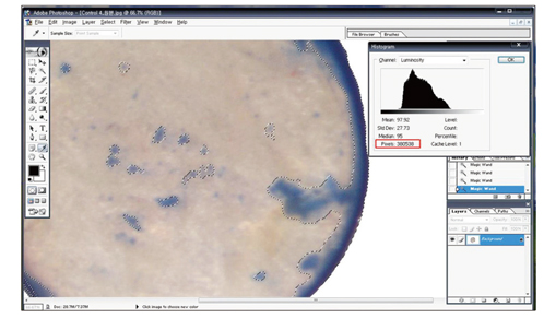
Fig. 1 Computer digitized planimetry using Photoshop ver. 9.0 was used to semi-quantitatively assess devitalized areas of endothelium. To determine damaged endothelial area percentages, we used pixel counts. The red box in the picture displays the pixel count in devitalized areas (gray).
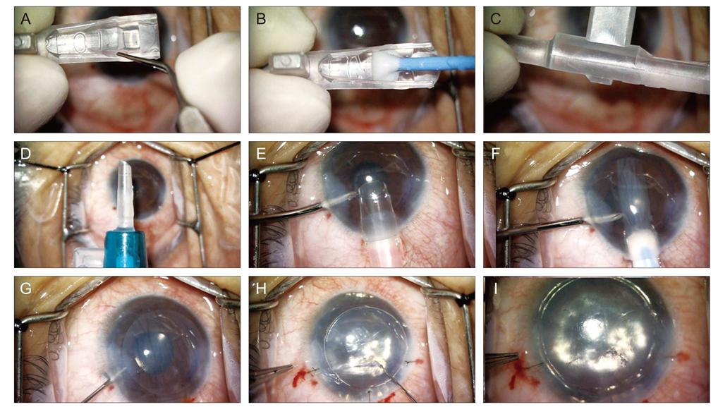
Fig. 2 The push-through insertion method using the Visian Implantable Collamer Lens (ICL) injector. (A) The donor lenticule was placed on the balanced salt solution filled ICL cartridge, (B) and then carefully advanced through the cartridge using the sponge plunger. (C) The donor lenticule was rolled not folded. (D) The photograph shows a cartridge mounted in the injector. (E) Photograph showing the injector inserted into the anterior chamber. (F) Photograph showing the donor lenticule being pushed into the anterior chamber. (G) Photograph showing unrolling of the donor lenticule as the injector is retracted. (H,I) Centration of the donor lenticule was performed through a venting stab incision.
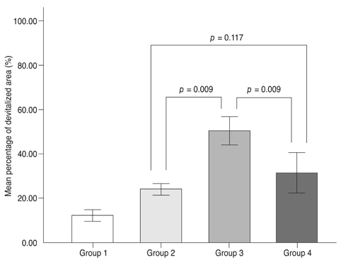
Fig. 3 Average endothelial cell damage percentages after the insertion of a donor lenticule in the four study groups. Group 1, control group (no delivery); group 2, Implantable Collamer Lens injector system-assisted delivery through a 3.2 mm limbal incision; group 3, forcep-assisted delivery through a 3.2 mm limbal incision; group 4, forcep-assisted delivery through a 4.0 mm limbal incision. Error bars: 95% confidence interval.
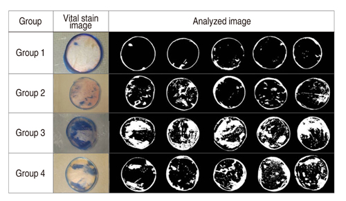
Fig. 4 Vital dye-stained corneal images and images analyzed using Adobe Photoshop. Group 1, control group (no delivery); group 2, Implantable Collamer Lens injector system-assisted delivery through a 3.2 mm limbal incision; group 3, forcep-assisted delivery through a 3.2 mm limbal incision; group 4, forcep-assisted delivery through a 4.0 mm limbal incision.
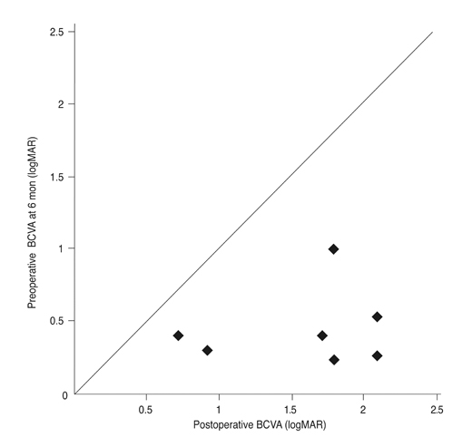
Fig. 5 Preoperative and postoperative best-corrected visual acuities (BCVA) at 6 months for all 7 patients, expressed as a logarithm of the minimum angle of resolution (logMAR).
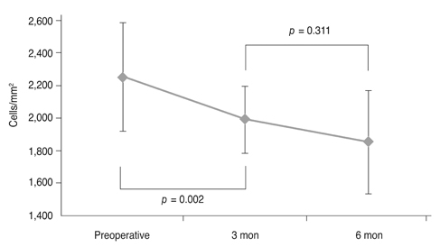
Fig. 6 Graph showing endothelial cell density as a function of time in 7 eyes grafted by Descemet's stripping endothelial keratoplasty.
Reference
-
1. Price FW Jr, Price MO. Descemet's stripping with endothelial keratoplasty in 50 eyes: a refractive neutral corneal transplant. J Refract Surg. 2005. 21:339–345.2. Chen ES, Terry MA, Shamie N, et al. Descemet-stripping automated endothelial keratoplasty: six-month results in a prospective study of 100 eyes. Cornea. 2008. 27:514–520.3. Gorovoy MS. Descemet-stripping automated endothelial keratoplasty. Cornea. 2006. 25:886–889.4. Melles GR, Wijdh RH, Nieuwendaal CP. A technique to excise the descemet membrane from a recipient cornea (descemetorhexis). Cornea. 2004. 23:286–288.5. Melles GR. Posterior lamellar keratoplasty: DLEK to DSEK to DMEK. Cornea. 2006. 25:879–881.6. Terry MA, Wall JM, Hoar KL, Ousley PJ. A prospective study of endothelial cell loss during the 2 years after deep lamellar endothelial keratoplasty. Ophthalmology. 2007. 114:631–639.7. Mehta JS, Por YM, Beuerman RW, Tan DT. Glide insertion technique for donor cornea lenticule during Descemet's stripping automated endothelial keratoplasty. J Cataract Refract Surg. 2007. 33:1846–1850.8. Kuo AN, Harvey TM, Afshari NA. Novel delivery method to reduce endothelial injury in descemet stripping automated endothelial keratoplasty. Am J Ophthalmol. 2008. 145:91–96.9. Busin M, Bhatt PR. Late detachment of donor graft after Descemet stripping automated endothelial keratoplasty. J Cataract Refract Surg. 2008. 34:159–160.10. Vajpayee RB, Agarwal T, Jhanji V, Sharma N. Modification in descemet-stripping automated endothelial keratoplasty: "Hitch suture" technique. Cornea. 2006. 25:1060–1062.11. Macsai MS, Kara-Jose AC. Suture technique for Descemet stripping and endothelial keratoplasty. Cornea. 2007. 26:1123–1126.12. Van Cleynenbreugel H, Hillenaar T, Remeijer L. Graft insertion during Descemet-stripping automated endothelial keratoplasty: pulling the graft inward. J Cataract Refract Surg. 2008. 34:534–536.13. Sarnicola V, Toro P. Descemet-stripping automated endothelial keratoplasty by using suture for donor insertion. Cornea. 2008. 27:825–829.14. Bethke W. Injecting innovation into DSEK. Rev Ophthalmol. 2008. 15:1.15. Ide T. Descemet's stripping automated endothelial keratoplasty injecting device. Expert Rev Ophthalmol. 2009. 4:5–9.16. Ide T, Yoo SH, Goldman JM, et al. Descemet-stripping automated endothelial keratoplasty: effect of inserting forceps on DSAEK donor tissue viability by using an in vitro delivery model and vital dye assay. Cornea. 2007. 26:1079–1081.17. Saad HA, Terry MA, Shamie N, et al. An easy and inexpensive method for quantitative analysis of endothelial damage by using vital dye staining and Adobe Photoshop software. Cornea. 2008. 27:818–824.18. Terry MA. Deep lamellar endothelial keratoplasty (DLEK): pursuing the ideal goals of endothelial replacement. Eye (Lond). 2003. 17:982–988.19. Bohringer D, Reinhard T, Spelsberg H, Sundmacher R. Influencing factors on chronic endothelial cell loss characterised in a homogeneous group of patients. Br J Ophthalmol. 2002. 86:35–38.20. Aralikatti A, Dean S, Busin M, Shah S. Pull-through technique for graft insertion in DSAEK. J Cataract Refract Surg. 2008. 34:341.21. Bradley JC, McCartney DL. Descemet's stripping automated endothelial keratoplasty in intraoperative floppy-iris syndrome: suture-drag technique. J Cataract Refract Surg. 2007. 33:1149–1150.22. Macaluso C. Closed-chamber pulling-injection system for donor graft insertion in endothelial keratoplasty. J Cataract Refract Surg. 2008. 34:353–356.23. Busin M, Bhatt PR, Scorcia V. A modified technique for descemet membrane stripping automated endothelial keratoplasty to minimize endothelial cell loss. Arch Ophthalmol. 2008. 126:1133–1137.24. Mehta JS, Por YM, Poh R, et al. Comparison of donor insertion techniques for descemet stripping automated endothelial keratoplasty. Arch Ophthalmol. 2008. 126:1383–1388.25. Terry MA, Saad HA, Shamie N, et al. Endothelial keratoplasty: the influence of insertion techniques and incision size on donor endothelial survival. Cornea. 2009. 28:24–31.
- Full Text Links
-
- Actions
-
Cited
- CITED
-
- Close
- Share
-
- Similar articles
-
- Long-term Clinical Outcomes of Implantable Collamer Lens
- Short-term Clinical Outcomes of Implantable Collamer Lens Implantation with Simultaneous Full Thickness Astigmatic Keratotomy
- Corneal Endothelial Changes After ICL (Implantable Collamer Lens) Insertion
- Descemet Membrane Endothelial Keratoplasty to Treat Graft Failure after Descemet Stripping Endothelial Keratoplasty
- Axial Length Change after Implantable Collamer Lens Implantation
