Korean J Ophthalmol.
2013 Feb;27(1):44-47. 10.3341/kjo.2013.27.1.44.
The Correlation of Differences in the Ocular Component Values with the Degree of Myopic Anisometropia
- Affiliations
-
- 1Department of Ophthalmology and Visual Science, The Catholic University of Korea College of Medicine, Seoul, Korea. yclee@cmcnu.or.kr
- KMID: 1501795
- DOI: http://doi.org/10.3341/kjo.2013.27.1.44
Abstract
- PURPOSE
To determine the relationship between the differences in the ocular component values with the degree of anisomyopia.
METHODS
Refraction, corneal power (CP), and biometry were examined in 50 myopic adults with refractive differences (RD) over 1.50 diopters (D). Ocular components were measured by ultrasound biometry and keratometry. The correlation between the differences in the ocular component values with the degree of anisomyopia was analyzed by linear regression analysis.
RESULTS
Among 50 adults with anisomyopia, 5 had RD from 1.50 to 2.99 D, 11 had RD from 3.00 to 3.99 D, 9 had RD from 4.00 to 5.99 D, 12 had RD from 6.00 to 7.99 D, 7 had RD from 8.00 to 11.99 D, and 6 had > or =12.00 D. There was no significant correlation between the ocular components (CP, crystalline lens thickness [LT], and anterior chamber depth [ACD], and the length from the cornea to the posterior surface of the lens [ACD + LT]) and the RD (p > 0.05). The RD showed a significantly positive correlation with vitreous chamber depth (VCD), and axial length (r = 0.963, p < 0.0001).
CONCLUSIONS
The severity of anisomyopia was not correlated with the between-eye differences in the anterior chamber values of the eye (CP, ACD, LT, ACD + LT). The severity of anisomyopia was significantly correlated with the between-eye differences in VCD.
Keyword
MeSH Terms
-
Adult
Aged
Anisometropia/complications/physiopathology/*ultrasonography
Anterior Chamber/*ultrasonography
Female
Follow-Up Studies
Humans
Male
Microscopy, Acoustic/methods
Middle Aged
Myopia/complications/physiopathology/*ultrasonography
Refraction, Ocular
Retrospective Studies
Severity of Illness Index
Young Adult
Figure
-

Fig. 1 Correlation between differences of vitreous chamber depth and refractive errors in anisomyopia. D = diopters.

Fig. 2 Correlation between differences of axial length and refractive errors in anisomyopia. D = diopters.
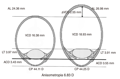
Fig. 3 A comparison of ocular components between the smaller eye and the larger eye in anisometropia group 6. AL = axial length; ΔVCD = difference of vitreous chamber depth between both eyes; VCD = vitreous chamber depth; LT = lens thickness; ACD = anterior chamber depth; CP = corneal power.
Cited by 1 articles
-
A Prospective Study of Anterior Segment Ocular Parameters in Anisometropia
Neha Singh, Jolly Rohatgi, Vinod Kumar
Korean J Ophthalmol. 2017;31(2):165-171. doi: 10.3341/kjo.2017.31.2.165.
Reference
-
1. Benjamin WJ, Borish IM. Borish's clinical refraction. 1998. Philadelphia: W. B. Saunders;2–17.2. Troilo D. Neonatal eye growth and emmetropisation: a literature review. Eye (Lond). 1992. 6(Pt 2):154–160.3. Olsen T, Arnarsson A, Sasaki H, et al. On the ocular refractive components: the Reykjavik Eye Study. Acta Ophthalmol Scand. 2007. 85:361–366.4. Wong TY, Foster PJ, Ng TP, et al. Variations in ocular biometry in an adult Chinese population in Singapore: the Tanjong Pagar Survey. Invest Ophthalmol Vis Sci. 2001. 42:73–80.5. Zaka-Ur-Rab S. Evaluation of relationship of ocular parameters and depth of anisometropic amblyopia with the degree of anisometropia. Indian J Ophthalmol. 2006. 54:99–103.6. Brown N. Lens changes with age and cataract: slit-image phothography. Symposium on the Human Lens in Relation to Cataract held at the Ciba Foundation. 1973. 1973 Jan 30-Feb 1; London, UK. Amsterdam: Elsevier;65–78.7. Larsen JS. The sagittal growth of the eye. 3. Ultrasonic measurement of the posterior segment (axial length of the vitreous) from birth to puberty. Acta Ophthalmol (Copenh). 1971. 49:441–453.8. Friedman NE, Mutti DO, Zadnik K. Corneal changes in schoolchildren. Optom Vis Sci. 1996. 73:552–557.9. Lin LL, Shih YF, Lee YC, et al. Changes in ocular refraction and its components among medical students: a 5-year longitudinal study. Optom Vis Sci. 1996. 73:495–498.10. Grosvenor T, Scott R. Three-year changes in refraction and its components in youth-onset and early adult-onset myopia. Optom Vis Sci. 1993. 70:677–683.11. Huynh SC, Wang XY, Ip J, et al. Prevalence and associations of anisometropia and aniso-astigmatism in a population based sample of 6 year old children. Br J Ophthalmol. 2006. 90:597–601.12. Patel VS, Simon JW, Schultze RL. Anisometropic amblyopia: axial length versus corneal curvature in children with severe refractive imbalance. J AAPOS. 2010. 14:396–398.13. Tayah D, Dall'coll MW, Alves MR. Refraction and its components in anisometropia. Arq Bras Oftalmol. 2009. 72:7–12.14. Lee SM, Kwon JY. Clinical study of anisometropia. J Korean Ophthalmol Soc. 2000. 41:2638–2644.
- Full Text Links
-
- Actions
-
Cited
- CITED
-
- Close
- Share
-
- Similar articles
-
- A Longitudinal Change of Spherical Equivalent in Anisometropic Children
- The Association Between Amblyopia and Anisometropia in Intermittent Exotropia
- The Amblyopia and Strabismus Accompanied with Anisometropia
- Relationship between Dominant Eye and Refractive Error in Myopic Anisometropia
- Clinical Features of Amblyopic Children with Myopic Anisometropia at a Tertiary Center
