Congenital Glaucoma from Sturge-Weber Syndrome: A Modified Surgical Approach
- Affiliations
-
- 1The Hong Kong Ophthalmic Associates, Central, Hong Kong.
- 2The Eye Institute, University of Hong Kong, Pokfulam, Hong Kong. ianyhwong@gmail.com
- KMID: 1499691
- DOI: http://doi.org/10.3341/kjo.2012.26.6.481
Abstract
- Sturge-Weber syndrome (SWS) is a rare congenital neurocutaneous disorder that causes congenital glaucoma. Previous experiences have shown that drainage procedures are often required to control associated glaucoma. The conventional surgical approach in trabeculectomy carries a significant risk of intraoperative expulsive hemorrhage. Here, we describe a modified approach of the conventional trabeculectomy technique, which may lower the risk of expulsive hemorrhage. A viscoelastic device was employed to maintain a steady intraocular pressure throughout the procedure. Details of the surgical technique and material used are described. One patient with congenital glaucoma associated with SWS underwent a successful trabeculectomy using the modified technique. Postoperative intraocular pressure was successfully reduced and no intraoperative complications occurred. We describe a successful case of trabeculectomy in a SWS case where a modified technique was applied.
Keyword
MeSH Terms
Figure
-

Fig. 1 (A) Clinical photo showing right facial haemangioma, characteristic of Sturge-Weber syndrome. (B) Right eye before primary operation showing features of bulphthalmos. (C) Normal left eye before the primary operation. (D) Operated eye one-year after operation, showing a well formed bleb superiorly.
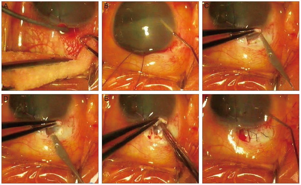
Fig. 2 Intraoperative photos. (A) Cauterisation of prominent episcleral vessels. (B) Injection of viscoelastics prior to anterior chamber penetration. (C) Appearance immediately prior to creating the inner window with slit knife. (D) Appearance of the inner window immediately after penetration with slit knife. Picture shows only a trace amount of viscoelastic egress from the anterior chamber. (E) Enlargement of inner window with a Kelly punch. (F) Testing aqueous outflow with the releasable suture tied.

Fig. 3 Post-operative intraocular pressure trend in the right eye. The intraocular pressure (IOP) decreased significantly after the primary trabeculectomy. Although there was a surge in the second week post-operatively, the IOP decreased to a lower level after the removal of the releasable suture on post-operative day 11. Pre-op = pre-operative.
Reference
-
1. Iwach AG, Hoskins HD Jr, Hetherington J Jr, Shaffer RN. Analysis of surgical and medical management of glaucoma in Sturge-Weber syndrome. Ophthalmology. 1990. 97:904–909.2. Keverline PO, Hiles DA. Trabeculectomy for adolescent onset glaucoma in the Sturge-Weber syndrome. J Pediatr Ophthalmol. 1976. 13:144–148.3. Bellows AR, Chylack LT Jr, Epstein DL, Hutchinson BT. Choroidal effusion during glaucoma surgery in patients with prominent episcleral vessels. Arch Ophthalmol. 1979. 97:493–497.4. Ali MA, Fahmy IA, Spaeth GL. Trabeculectomy for glaucoma associated with Sturge-Weber syndrome. Ophthalmic Surg. 1990. 21:352–355.5. Audren F, Abitbol O, Dureau P, et al. Non-penetrating deep sclerectomy for glaucoma associated with Sturge-Weber syndrome. Acta Ophthalmol Scand. 2006. 84:656–660.6. Taylor DM. Expulsive hemorrhage. Am J Ophthalmol. 1974. 78:961–966.7. Eibschitz-Tsimhoni M, Lichter PR, Del Monte MA, et al. Assessing the need for posterior sclerotomy at the time of filtering surgery in patients with Sturge-Weber syndrome. Ophthalmology. 2003. 110:1361–1363.8. Lane D, Motolko M, Yan DB, Ethier CR. Effect of Healon and Viscoat on outflow facility in human cadaver eyes. J Cataract Refract Surg. 2000. 26:277–281.9. Wu SC, Huang SC, Kuo CL, et al. Reversal of optic disc cupping after trabeculotomy in primary congenital glaucoma. Can J Ophthalmol. 2002. 37:337–341.
- Full Text Links
-
- Actions
-
Cited
- CITED
-
- Close
- Share
-
- Similar articles
-
- A Case of Sturge-Weber Syndrome
- Sturge-Weber Syndrome with Congenital Ocular Anomaly
- Clinical Experience of Treatment in a Case of Sturge-Weber Syndrome with Bilateral Glaucoma
- Anesthesia for a Sturge-Weber Syndrome Patient with Severe Facial Hemangioma: A case report
- Clinical Features and Surgical Outcomes of Sturge-Weber Syndrome with Glaucoma
