Intervention Using Ultrasonography
- Affiliations
-
- 1Department of Orthopedic Surgery, St. Paul's Hospital, The Catholic University of Korea School of Medicine, Seoul, Korea. hssongmd@yahoo.com
- KMID: 1494146
- DOI: http://doi.org/10.4055/jkoa.2013.48.5.342
Abstract
- Intervention using ultrasonography includes injection, aspiration, needling (barbotage), nerve block, tumor biopsy, and removal of the foreign body. The principles of disinfection during skin preparation, as well as handling of the transducer and needle should be maintained. The needle could be visualized on the ultrasonographic image, and could be tracked during the intervention. Factors affecting visualization of the needle on ultrasonography include the diameter and the incidence angle of the needle. Ultrasonography can be classified as an indirect technique or a real-time technique according to constant use. According to the angle between the transducer and needle, it can be classified as a lateral approach or a coaxial approach.
Keyword
MeSH Terms
Figure
-

Figure 1 Sterile envelope for the transducer: (A) sterile-packed and (B) wrapped and rubber-banded.
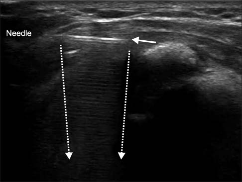
Figure 2 Ultrasonography shows the reverberation by the needle (arrow). Dotted arrows indicate the comet-tail artifact behind the needle.

Figure 3 Long needle with a stylet.
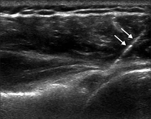
Figure 4 Ultrasonography of the posterior approach to the shoulder joint shows no reverberation by the needle. Arrows indicate the needle.

Figure 5 Incidence angle of the needle can be increased by change of the puncture site apart from the transducer: (A) near the transducer, (B) apart from the transducer.

Figure 6 Incidence angle of the needle can be increased by pressing on the opposite side of the transducer: (A) before, (B) after the heel-toe maneuver.
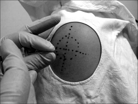
Figure 7 The indirect technique. Two perpendicular lines crossing the center of the lesion are marked with dotted lines.
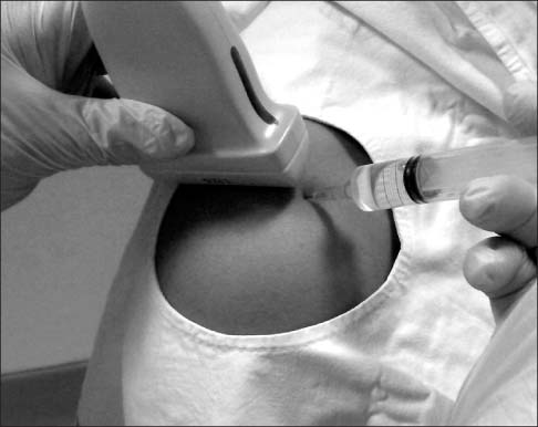
Figure 8 Lateral approach (in-plane technique) for injection into the aromioclavicular joint. The needle is aligned with the small side of the transducer.

Figure 9 Coaxial approach (out-of-plane technique) for injection into the aromioclavicular joint. The needle is aligned with the broad side of the transducer.
Reference
-
1. Douglas BR, Charboneau JW, Reading CC. Ultrasound-guided intervention: expanding horizons. Radiol Clin North Am. 2001; 39:415–428.2. Lento PH, Strakowski JA. The use of ultrasound in guiding musculoskeletal interventional procedures. Phys Med Rehabil Clin N Am. 2010; 21:559–583.
Article3. Zwar RB, Read JW, Noakes JB. Sonographically guided glenohumeral joint injection. AJR Am J Roentgenol. 2004; 183:48–50.
Article4. Chiou HJ, Chou YH, Wu JJ, et al. Alternative and effective treatment of shoulder ganglion cyst: ultrasonographically guided aspiration. J Ultrasound Med. 1999; 18:531–535.
Article5. Aina R, Cardinal E, Bureau NJ, Aubin B, Brassard P. Calcific shoulder tendinitis: treatment with modified US-guided fine-needle technique. Radiology. 2001; 221:455–461.
Article6. Ootaki C, Hayashi H, Amano M. Ultrasound-guided infraclavicular brachial plexus block: an alternative technique to anatomical landmark-guided approaches. Reg Anesth Pain Med. 2000; 25:600–604.
Article7. Torriani M, Etchebehere M, Amstalden E. Sonographically guided core needle biopsy of bone and soft tissue tumors. J Ultrasound Med. 2002; 21:275–281.
Article8. Mizel MS, Steinmetz ND, Trepman E. Detection of wooden foreign bodies in muscle tissue: experimental comparison of computed tomography, magnetic resonance imaging, and ultrasonography. Foot Ankle Int. 1994; 15:437–443.
Article9. Bianchi S, Zamorani MP. US-guided interventional procedures. In : Bianchi S, Zamorani MP, editors. Ultrasound of the musculoskeletal system. New Delhi: Springer Berlin Heidelberg;2007. p. 891–917.10. Saker MB, Kane RA, Matalon TAS. Factors affecting and techniques to improve needle visualization. Semin intervent Radiol. 1997; 14:471–475.
Article11. Bisceglia M, Matalon TA, Silver B. The pump maneuver: an atraumatic adjunct to enhance US needle tip localization. Radiology. 1990; 176:867–868.
Article
- Full Text Links
-
- Actions
-
Cited
- CITED
-
- Close
- Share
-
- Similar articles
-
- Re: Clinical significance of isolated macrocalcifications detected by ultrasonography
- Usefulness of musculoskeletal ultrasonography for treatment of shoulder pain
- ULTRASONOGRAPHY is now in SCIE
- ULTRASONOGRAPHY: how to raise the impact factor
- ULTRASONOGRAPHY: the fifth anniversary of its global re-launch
