Intraosseous Arteriovenous Malformation of the Sphenoid Bone Presenting with Orbital Symptoms Mimicking Cavernous Sinus Dural Arteriovenous Fistula: A Case Report
- Affiliations
-
- 1Department of Neurosurgery, Ulsan University Hospital, University of Ulsan College of Medicine, Ulsan, Korea.
- 2Department of Neurosurgery, College of Medicine, Yeungnam University, Daegu, Korea.
- 3Department of Neurosurgery, College of Medicine, Dankook University, Cheonan, Korea.
- 4Department of Neurosurgery, Asan Medical Center, University of Ulsan College of Medicine, Seoul, Korea.
- 5Department of Radiology, Asan Medical Center, University of Ulsan College of Medicine, Seoul, Korea. dhlee@amc.seoul.kr
- KMID: 1491448
- DOI: http://doi.org/10.7461/jcen.2013.15.3.251
Abstract
- Intraosseous arteriovenous malformation (AVM) in the craniofacial region is rare. When it occurs, it is predominantly located in the mandible and maxilla. We encountered a 43-year-old woman with Klippel-Trenaunay syndrome affecting the right lower extremity who presented with a left orbital chemosis and proptosis mimicking the cavernous sinus dural arteriovenous fistula. Computed tomography angiography revealed an intraosseous AVM of the sphenoid bone. The patient's symptoms were completely relieved after embolization with Onyx. We report an extremely rare case of intraosseous AVM involving the sphenoid bone, associated with Klippel-Trenaunay syndrome.
Keyword
MeSH Terms
Figure
-
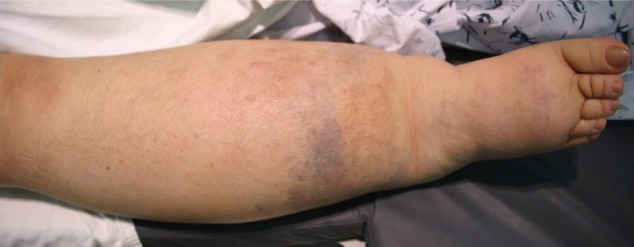
Fig. 1 Photography shows the hypertrophy of our patient's right lower extremity, with bluish cutaneous stains.
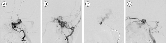
Fig. 2 (A, B) Left external carotid angiogram shows the arteriovenous malformation (AVM) fed by the internal maxillary and accessory meningeal arteries. (C) Left accessory meningeal artery injection demonstrates a high flow AVM. (D) The nidus is drained via the ipsilateral cavernous sinus with significant reflux into the ipsilateral superior and inferior ophthalmic veins while the inferior petrosal sinus is patent.
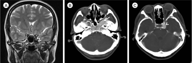
Fig. 3 (A) Brain magnetic resonance image with T2-weighted coronal image shows that the vascular mass corresponded with the dark-signaled mass (Double arrowheads) in the left side of the sphenoid body and the medial aspect of the greater wing. (B, C) Contrast-enhanced brain computed tomography shows a vascular abnormality localized in the sphenoid bone near the left paracavernous region. The abnormal vascular lesion (Arrow) is mainly located in sphenoid bone. The draining vein (Arrowhead) is located in the paracavernous region.
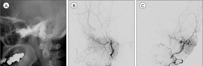
Fig. 4 Three-month follow-up angiogram shows complete obliteration of the AVM. (A) Scout image. (B) Left external carotid angiogram, lateral view of arterial phase. (C) Left external carotid angiogram, anteroposterior view of arterial phase.
Reference
-
1. Alomari AI, Orbach DB, Mulliken JB, Bisdorff A, Fishman SJ, Norbash A, et al. Klippel-Trenaunay syndrome and spinal arteriovenous malformations: An erroneous association. AJNR Am J Neuroradiol. 2010; 10. 31(9):1608–1612. PMID: 20651014.
Article2. Fan X, Qiu W, Zhang Z, Mao Q. Comparative study of clinical manifestation, plain-film radiography, and computed tomographic scan in arteriovenous malformations of the jaws. Oral Surg Oral Med Oral Pathol Oral Radiol Endod. 2002; 10. 94(4):503–509. PMID: 12374928.
Article3. Jacob AG, Driscoll DJ, Shaughnessy WJ, Stanson AW, Clay RP, Gloviczki P. Klippel-Trenaunay syndrome: Spectrum and management. Mayo Clin Proc. 1998; 1. 73(1):28–36. PMID: 9443675.
Article4. Jung C, Kwon BJ, Kwon OK, Baik SK, Han MH, Kim JE, et al. Intraosseous cranial dural arteriovenous fistula treated with transvenous embolization. AJNR Am J Neuroradiol. 2009; 6. 30(6):1173–1177. PMID: 19246532.
Article5. Knych SA, Goldberg MJ, Wolfe HJ. Intraosseous arteriovenous malformation in a pediatric patient. Clin Orthop Relat Res. 1992; 3. (276):307–312. PMID: 1537171.6. Louis RG Jr, Yen CP, Mohila CA, Mandell JW, Sheehan J. A rare intraosseous arteriovenous malformation of the spine. J Neurosurg Spine. 2011; 9. 15(3):336–339. PMID: 21663404.
Article7. Malik GM, Mahmood A, Mehta BA. Dural arteriovenous malformation of the skull base with intraosseous vascular nidus. Report of two cases. J Neurosurg. 1994; 10. 81(4):620–623. PMID: 7931600.8. Oduber CE, van der Horst CM, Hennekam RC. Klippel-Trenaunay syndrome: Diagnostic criteria and hypothesis on etiology. Ann Plast Surg. 2008; 2. 60(2):217–223. PMID: 18216519.9. Star A, Fuller CE, Landas SK. Intracranial aneurysms in klippel-trenaunay/weber syndromes: Case report. Neurosurgery. 2010; 5. 66(5):E1027–E1028. discussion E1028. PMID: 20404675.
- Full Text Links
-
- Actions
-
Cited
- CITED
-
- Close
- Share
-
- Similar articles
-
- Endovascular Treatment of Dural Sinus Malformation in Infant: A Case Report
- A Case of Dural Arterio-venous Malformation Involving the Cavernous Sinus
- Endovascular management of cavernous sinus dural arteriovenous fistulas: Overall review and considerations
- A Case of Unilateral Exophthalmos Caused by a Dural Arteriovenous Malformation in Thyroid-Associated Ophthalmopathy
- Occurrence of De Novo Dural Arteriovenous Fistula after Transvenous Embolization of Dural Arteriovenous Fistula : Case Reports of Two Patients
