Valsalva Aneurysm Filled with Thrombi Mimicking a Cardiac Tumor
- Affiliations
-
- 1Division of Cardiology, Osaka Rosai Hospital, Osaka, Japan. mnishino@orh.go.jp
- KMID: 1491107
- DOI: http://doi.org/10.4070/kcj.2012.42.12.869
Abstract
- A Valsalva aneurysm filled with thrombi can be difficult to diagnose, because it mimics a cardiac tumor. Both cardiac magnetic resonance imaging (MRI) and transesophageal echocardiogram (TEE) were performed on a patient who showed a low-echoic mass located between the atrial septum and the non-coronary sinus. Based on MRI findings allowing tissue characterization and the accurate location of the mass and the TEE findings of an irregular surface of the mass and a partial defect in the edge of the non-coronary sinus, we diagnosed the mass as a thrombosed Valsalva aneurysm that had perforated the inter-atrial septum. The operative findings coincided with the preoperative diagnosis. Both MRI and TEE are useful for diagnosing this condition.
MeSH Terms
Figure
-
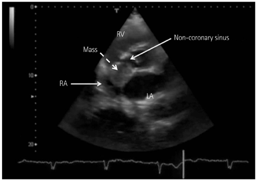
Fig. 1 The transthoracic echocardiography demonstrates a low echoic mass (26×30 mm) (dotted arrow) located between the atrial septum and the noncoronary sinus. LA: left atrium, RA: right atrium, RV: right ventricle.
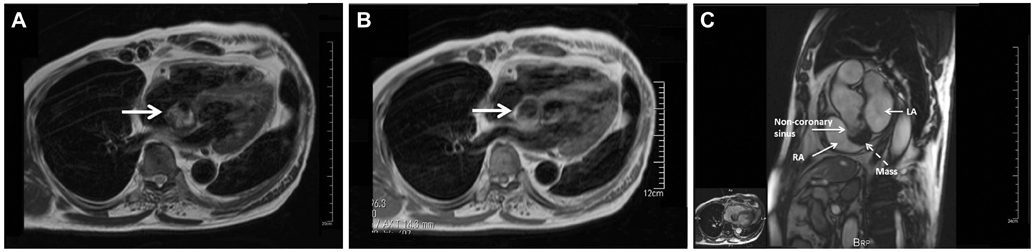
Fig. 2 The findings from magnetic resonance imaging (MRI). A: T2-weighted fast spin-echo image. The arrow shows the mass, which presented with high signal intensity mostly, mixed with focal low signal intensity on T2-weighted fast spin-echo image. B: T1-weighted fast spin-echo image. The arrow shows the mass, which presented with dark signal intensity. C: a cine MRI reveals that the mass is connected to the non-coronary sinus and is not located in the right atrium (RA). LA: left atrium.
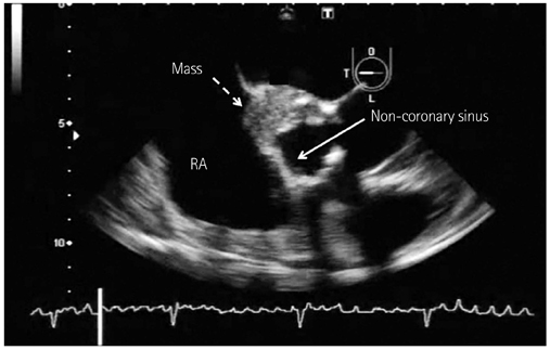
Fig. 3 A transesophageal echocardiography (TEE) which shows a dilated non-coronary sinus filled with a low echoic mass suspected to be thrombi (dotted arrow), because the mass surface in the non-coronary sinus is irregular. The edge of the non-coronary sinus is defected, which might have indicated a perforated lumen. LA: left atrium, RA: right atrium.
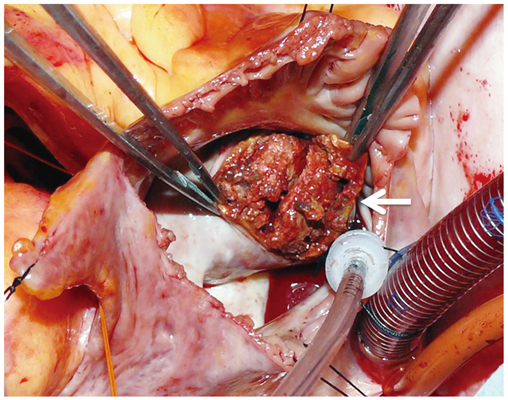
Fig. 4 The operative findings reveal that the non-coronary sinus is defected and filled with thrombi (arrow).
Reference
-
1. Flynn MS, Castello R, McBride LW, Labovitz AJ. Ruptured congenital aneurysm of the sinus of Valsalva with persistent left superior vena cava imaged by intraoperative transesophageal echocardiography. Am Heart J. 1993. 125:1185–1187.2. Copeland JG, Valdes-Cruz L, Sahn DJ. Endomyocardial biopsy with fluoroscopic and two-dimensional echocardiographic guidance: case report of a patient suspected of having multiple cardiac tumors. Clin Cardiol. 1984. 7:449–452.3. Meier JH, Seward JB, Miller FA Jr, Oh JK, Enriquez-Sarano M. Aneurysms in the left ventricular outflow tract: clinical presentation, causes, and echocardiographic features. J Am Soc Echocardiogr. 1998. 11:729–745.4. Popescu BA, Muraru D, Beladan CC, Lăcău IS, Ginghină C. Images in cardiology: atrioventricular block in the elderly: does echocardiography hold the key? J Am Coll Cardiol. 2011. 57:219.5. Wang ZJ, Zou CW, Li DC, et al. Surgical repair of sinus of Valsalva aneurysm in Asian patients. Ann Thorac Surg. 2007. 84:156–160.6. Nagueh SF. Assessment of valvular regurgitation with Doppler echocardiography. Cardiol Clin. 1998. 16:405–419.7. Mookadam F, Haley J, Mendrick E. Rare cause of right heart failure: contained rupture of a sinus of Valsalva aneurysm associated intraventricular septal aneurysm. Eur J Echocardiogr. 2005. 6:221–224.8. Lijoi A, Parodi E, Passerone GC, Scarano F, Caruso D, Iannetti MV. Unruptured aneurysm of the left sinus of valsalva causing coronary insufficiency: case report and review of the literature. Tex Heart Inst J. 2002. 29:40–44.9. Shahrabani RM, Jairaj PS. Unruptured aneurysm of the sinus of Valsalva: a potential source of cerebrovascular embolism. Br Heart J. 1993. 69:266–267.10. Sasaki S, Asano M, Fukuda K, et al. Unruptured sinus of Valsalva aneurysm suspected to be a cardiac tumor. Ann Thorac Surg. 2009. 87:1619.11. Hoey ET, Kanagasingam A, Sivananthan MU. Sinus of valsalva aneurysms: assessment with cardiovascular MRI. AJR Am J Roentgenol. 2010. 194:W495–W504.
- Full Text Links
-
- Actions
-
Cited
- CITED
-
- Close
- Share
-
- Similar articles
-
- Huge Aneurysm of the Sinus of Valsalva Compressing the Left Atrium
- A Rare Case of Unruptured Sinus of Valsalva Aneurysm Obstructing the Right Ventricular Outflow Tract
- A Case of Perimembranous Ventricular Septal Defect Associated with Sinus of Valsalva Aneurysm Mimicking Membranous Septal Aneurysm
- Acute Myocardial Infarction Due to an Unruptured Sinus of Valsalva Aneurysm in a Patient with Behcet's Syndrome
- A Case of Ruptured Aneurysm of the Sinus of Valsalva
