Clin Exp Otorhinolaryngol.
2012 Dec;5(4):234-236. 10.3342/ceo.2012.5.4.234.
A Posterior Petrous Meningioma with Recurrent Vertigo
- Affiliations
-
- 1Department of Otorhinolaryngology-Head and Neck Surgery, Konyang University College of Medicine, Daejeon, Korea.
- 2Department of Otolaryngology, Ajou University School of Medicine, Suwon, Korea. yhc@ajou.ac.kr
- KMID: 1489085
- DOI: http://doi.org/10.3342/ceo.2012.5.4.234
Abstract
- Meningioma's account for around 15% of all primary brain tumors with some 10% of meningiomas arising in the posterior fossa. In rare cases, a meningioma can form around the endolymphatic sac. When formed in the posterior fossa, meningioma tumors can produce vague, non-specific vertiginous symptoms. Research has observed that a subset of these lesions could produce symptoms indistinguishable from those of Meniere's disease. Therefore, we described the clinical features of a case of posterior petrous meningioma with recurrent vertigo as well as the substantial resolution of symptoms after tumor removal via transmastoid approach.
Keyword
Figure
-

Fig. 1 A preoperative temporal bone computed tomography scan shows a round bony defect in the left posterior petrous region (arrow).
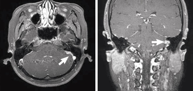
Fig. 2 A gadolinium enhanced temporal magnetic resonance imaging scan shows a homogenous enhanced mass in the left posterior petrous region (arrow).
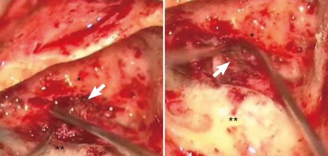
Fig. 3 Tumor was found to be attached to the dura overlying the endolymphatic sac and resected via a transmastoid approach (arrow).*Posterior semicircular canal. **Singmoid sinus.
Reference
-
1. Brackmann DE, Bartels LJ. Rare tumors of the cerebellopontine angle. Otolaryngol Head Neck Surg. 1980; Sep-Oct. 88(5):555–559. PMID: 6969383.
Article2. Coelho DH, Roland JT Jr, Golfinos JG. Posterior fossa meningiomas presenting with Ménière's-like symptoms: case report. Neurosurgery. 2008; 11. 63(5):E1001. PMID: 19005363.3. Maniglia AJ. Intra and extracranial meningiomas involving the temporal bone. Laryngoscope. 1978; 9. 88(9 Pt 2):Suppl 12. 1–58. PMID: 355750.
Article4. Scott M. The surgical management of meningiomas of the cerebellar fossa. Surg Gynecol Obstet. 1972; 10. 135(4):545–550. PMID: 5077720.5. Castellano F, Ruggiero G. Meningiomas of the posterior fossa. Acta Radiol Suppl. 1953; 104:1–177. PMID: 13147998.6. Laird FJ, Harner SG, Laws ER Jr, Reese DF. Meningiomas of the cerebellopontine angle. Otolaryngol Head Neck Surg. 1985; 4. 93(2):163–167. PMID: 3921906.
Article7. Friedman RA, Nelson RA, Harris JP. Posterior fossa meningiomas intimately involved with the endolymphatic sac. Am J Otol. 1996; 7. 17(4):612–616. PMID: 8841708.8. Cmejrek RC, Megerian CA. Obstructing lesions of the endolymphatic sac and duct mimicking Ménière's disease. Ear Nose Throat J. 2004; 11. 83(11):753–756. PMID: 15628631.
Article9. Hadley MN, Grahm TW, Daspit CP, Spetzler RF. Otolaryngologic manifestations of posterior fossa arachnoid cysts. Laryngoscope. 1985; 6. 95(6):678–681. PMID: 3999903.
Article10. Haberkamp TJ, Monsell EM, House WF, Levine SC, Piazza L. Diagnosis and treatment of arachnoid cysts of the posterior fossa. Otolaryngol Head Neck Surg. 1990; 10. 103(4):610–614. PMID: 2123320.
Article11. Kimura RS, Schuknecht HF. Membranous hydrops in the inner ear of the guinea pig after obliteration of the endolymphatic sac. Pract Otorhinolaryngol (Basel). 1965; 27(6):343–354.
Article
- Full Text Links
-
- Actions
-
Cited
- CITED
-
- Close
- Share
-
- Similar articles
-
- A Case of Posterior Fossa Meningioma Mimicking Meniere's Disease
- A Case of Jugular Foramen Clear Cell Meningioma Presented as Sensorineural Hearing Loss and Recurrent Vertigo
- A Case of Posterior Fossa Meningioma Presenting with Meniere's-Like Symptoms
- Diagnosis of Benign Paroxysmal Positional Vertigo
- Malignant(papillary) Meningioma of the Petrous Bone Extending to Cerebellopontine Angle and Infratemporal Fossa: Case Report
