Korean J Phys Anthropol.
2013 Sep;26(3):101-104. 10.11637/kjpa.2013.26.3.101.
Frequency and Quantity of the C7 Contribution to the Ulnar Nerve
- Affiliations
-
- 1Department of Anatomy, Kwandong University College of Medicine, Korea. kslee@kwandong.ac.kr
- KMID: 1488820
- DOI: http://doi.org/10.11637/kjpa.2013.26.3.101
Abstract
- Many anatomists and clinicians who investigate the peripheral nerve concern about the composition of the spinal roots of each terminal nerve of the brachial plexus. From this viewpoint, the spinal root composition of the ulnar nerve is still unclear. Several anatomy textbooks describe that the ulnar nerve is composed of the ventral rami of the C8, T1 and often C7. There is no literature regarding the frequency and contribution quantity of C7 to the ulnar nerve. The purpose of present study was to determine frequency and contribution quantity of the C7 to the ulnar nerve. Fifty cadavers of brachial plexus were obtained from cadavers of Korean adults. The brachial plexus containing the ulnar nerve were extracted from the axilla and the extracted samples were immersed in Guanidine-HCl (0.2 M) for 2 weeks to soften the connective tissue around the nerve bundles. C7 was contributed to the ulnar nerve in all sides (100%). The numbers of the myelinated axons of C7 participating to the ulnar nerve was 1,452+/-429 (mean+/-S.D.). Thus the C7 can be considered as always participating component of the ulnar nerve, not often participation, although numbers of the myelinated axons of C7 was lesser than those of the C8, but similar to those of the T1. The results of the study provide a reference for accurate diagnosis and treatment regarding ulnar nerve injury due to various accidents.
Keyword
MeSH Terms
Figure
-
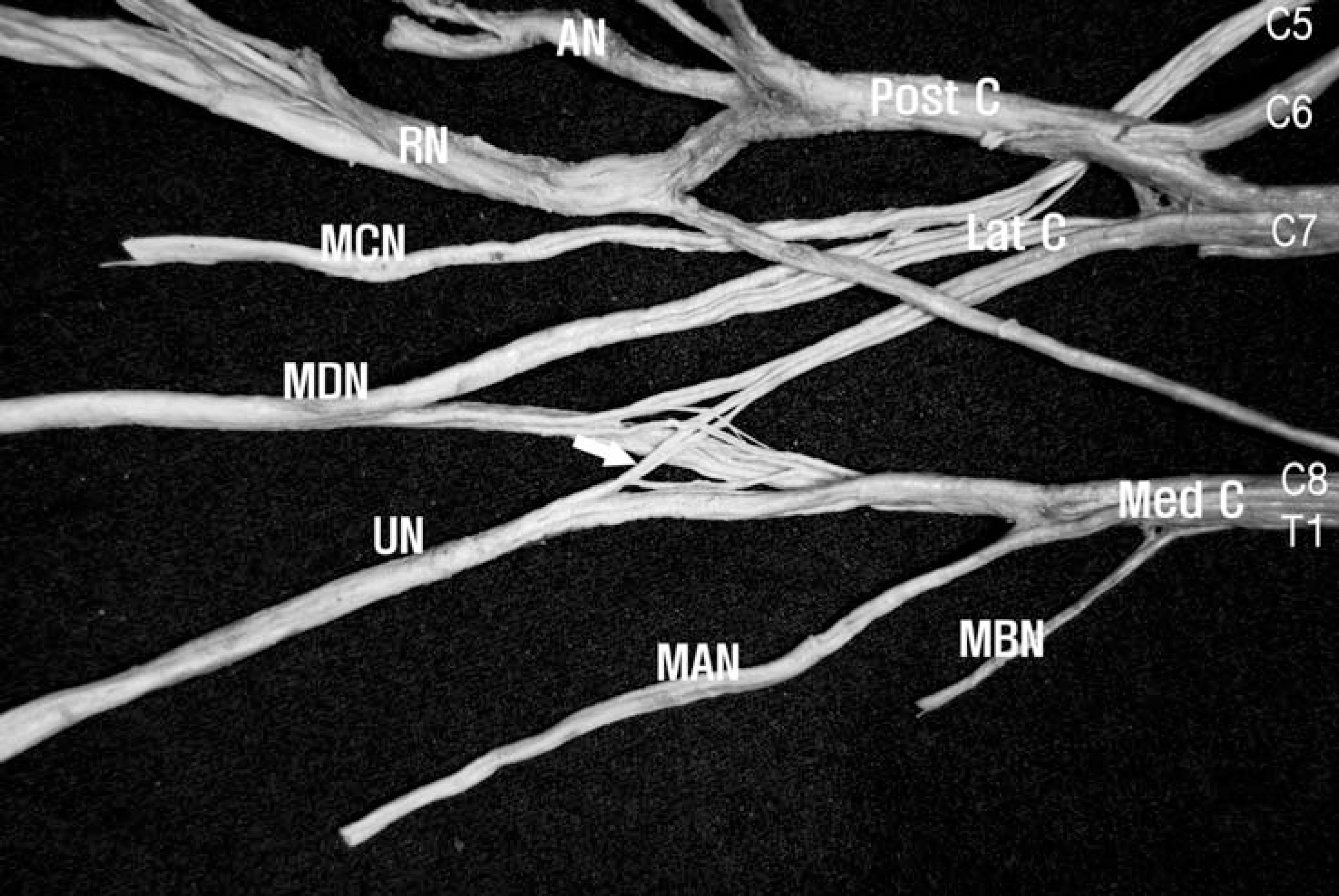
Fig. 1. A photograph showing the C7 fascicles(arrow) constituting the ulnar nerve in the posterior aspect of the brachial plexus. C7 fasci-cles constituting the ulnar nerve branched out at the distal portion of the lateral cord. AN, axillary nerve, Lat C, lateral cord; MAN, medial antebrachial cutaneous nerve; MBN, medial brachial cutaneous nerve; MCN, musculocutaneous nerve; MDN, median nerve; Med C, medial cord; Post C, posterior cord; RN, radial nerve; UN, ulnar nerve.
Reference
-
References
1. Romanes GJ. Cunningham's Textbook of Anatomy. 12th ed.New York: Oxford University Press;1981. p. 328–9.2. Ellis H. Clinical Anatomy-A revision and applied anatomy for clinical students. 8th ed.London: Blackwell Scientific Publications;1992. p. 209.3. Moore KL, Dalley AF. Clinically Oriented Anatomy. 5th ed.Baltimore: Lippincott Willams & Wilkins;2006. p. 777.4. Standring S. Gray's Anatomy. 39th ed.Edinburgh: Elsevier Churchill Livingston;2005. p. 847, 877.5. Fuss FK. The lateral root of the ulnar nerve. Acta Anat (Basel). 1989; 134:199–205.6. Pyun SB, Kang S, Kwon HK. Anatomical and electrophysiological myotome corresponding to the flexor carpi ulnaris muscle. J Korean Med Sci. 2010; 25:454–57.7. Norkus T, Norkus M, Ramanauskas T. Donor, recipient and nerve grafts in brachial reconstruction: anatomical and technical features of facilitating the exposure. Surg Radiol Anat. 2005; 27:524–30.8. Lindner HH. Clinical Anatomy. International ed. London: Appleton & Lange;1989. p. 554.9. MacKinon PCB, Morris JF. Oxford Textbook of Functional Anatomy. MusculoSkeletal System(Vol. 1). 2nd ed.Oxford: Oxford University Press;2005. p. 85.10. Russell SM. Examination of Peripheral Nerve Injuries. 1st ed.New York: Thieme;2006. p. 22–7.11. Dumitru D, Zwarts M. Brachial plexopathies and proximal mononeuropathies. Dumitru M, Amato A, Zwarts M, editors. editors.Electrodiagnostic Medicine. 2nd ed.Philadelphia: Hanley & Belfus;2002. p. 777–836. Cited from Pyun et al.(2010).
Article12. Hur MS, Lee KS. Quantification of the nerve fiber of the terminal branches of the typical brachial plexus. Korean J Phys Anthropol. 2011; 24:135–40.
Article
- Full Text Links
-
- Actions
-
Cited
- CITED
-
- Close
- Share
-
- Similar articles
-
- Quantification of the Nerve Fiber of the Terminal Branches of the Typical Brachial Plexus
- Tardy Ulnar Nerve Palsy with Recurrent Ulnar-Nerve Dislocation: Case Report
- An Unusual Communication between the Radial and the Ulnar Nerves
- Ulnar Neuropathy Caused by a Schwannoma in the Guyon's Cannal
- Anatomic Variations of the Spinal Origins of the Main Terminal Branches of the Brachial Plexus
