Analysis of the Development of the Nasal Septum according to Age and Gender Using MRI
- Affiliations
-
- 1Department of Otorhinolaryngology-Head & Neck Surgery, College of Medicine, Dankook University, Cheonan, Korea. docjung@paran.com
- KMID: 1486033
- DOI: http://doi.org/10.3342/ceo.2008.1.1.29
Abstract
OBJECTIVES
This study was designed to evaluate the normal development of the nasal septum in Koreans using sagittal MRI for the valuable clinical information on septal procedures. METHODS: Two hundred eighty patients who had their whole nasal septum visualized in the midline sagittal view were selected among the 3,904 patients with brain MRI from January, 2004 to December, 2006 at Dankook University Hospital. The patients who had a history of nasal septal surgery or nasal trauma were excluded. Following parameters are calculated and analyzed: lengths of bony and cartilage dorsum and septal cartilage-nasal bone overlap, total septal area, septal cartilage area and, the proportion of the cartilage area to septal area and the maximal harvestable cartilage for grafting were calculated using the PAC(TM) program. RESULTS: All the parameters were increased until adolescence. Thereafter, bony dorsal length, cartilage dorsal length, total dorsal length, total septal area and maximal harvestable cartilage for grafting have not changed significantly with age, while SC-NB overlap length, septal cartilage area, and proportion of the cartilage area to the total septal area were significantly decreased with age. The SC-NB overlap length was positively correlated with the septal cartilage area and the proportion of the cartilage area to the total septal area. CONCLUSION: The small septal cartilage area and its proportion to the total septal area were significantly correlated with a short overlap length of the septal cartilage under the nasal bone. Septal procedures should be carefully performed in the elderly due to the risk of incurring saddle nose.
Keyword
MeSH Terms
Figure
-

Fig. 1 Measurement of the length and area of the septum. (A) The bony dorsal length (from the most posterior point in the bony nasofrontal angle to the rhinion), (B) The cartilage dorsal length (from the rhinion to the imaginary anterior septal angle), (C) The SC-NB overlap length (from the rhinion to the most posterior point of the septal cartilage contacting the nasal bone), (D) The total septal area, (E) The septal cartilage area, (F) The maximal area for grafting. SC: septal cartilage; NB: nasal bone.
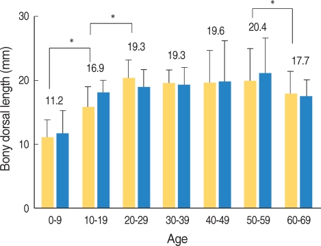
Fig. 2 Changes of the bony dorsal length according to age and gender. The bony dorsal length was increased until the 3rd decade and then it was not significantly changed with age. There was no significant gender difference. The diagonally stripped bars indicate males, the open bars the females. *P<0.05.
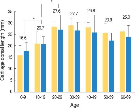
Fig. 3 Changes of the cartilage dorsal length according to age and gender. The cartilaginous dorsal length was increased until the 3rd decade and then it was not changed significantly with age. There was no significant gender difference. The diagonally stripped bars indicate the males and the open bars the females. *P<0.05.

Fig. 4 Changes of the total dorsal length according to age and gender. The total dorsal length was increased until the 3rd decade and then it was not changed significantly with age. There was no significant gender difference. The diagonally stripped bars indicate the males and the open bars the females. *P<0.05.
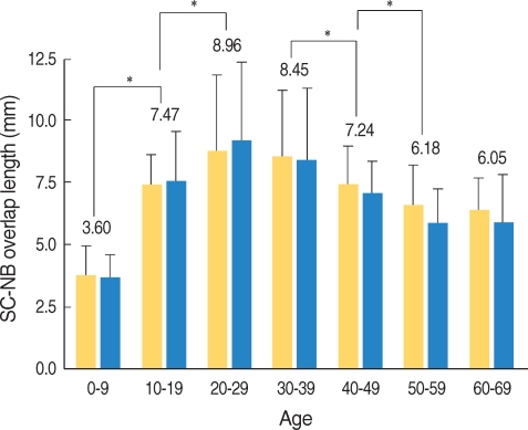
Fig. 5 Changes of the septal cartilage-nasal bone overlap length according to age and gender. The septal cartilage-nasal bone overlap length was increased until the 3rd decade and then it was decreased significantly with age. There was no significant gender difference.SC: septal cartilage; NB: nasal bone. The diagonally stripped bars indicate the males and the open bars the females. *P<0.05.
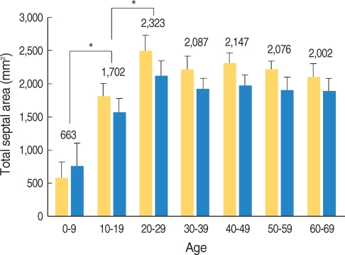
Fig. 6 Changes of the total septal area according to age and gender. The total septal area was increased until the 3rd decade and then it was not changed significantly with age. There was no significant gender difference. The diagonally stripped bars indicate the males and the open bars the females. *P<0.05.

Fig. 7 Changes of the septal cartilage area according to age and gender. The septal cartilage area was increased until the 3rd decade and then it was decreased significantly with age. There was no significant gender difference. The diagonally stripped bars indicate the males and the open bars the females. *P<0.05.

Fig. 8 Changes of the proportion of the cartilage area according to age and gender. The proportion of the cartilage area was decreased significantly with age. Solid lines indicate males, the dotted lines the females.

Fig. 9 Changes of the maximal available cartilage for grafting according to age and gender. The maximal available cartilage for grafting was increased until the 3rd decade and then it was decreased significantly with age. There was no significant gender difference. Diagonally stripped bars indicate the males, the open bars the females. *P<0.05.
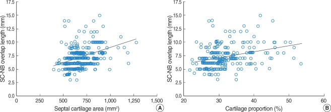
Fig. 10 (A) Correlation between the SC-NB overlap length and the septal cartilage area (r=0.386, P<0.001), (B) Correlation between the SCNB overlap length and the proportion of the cartilage area (r=0.327, P<0.001). SC, septal cartilage; NB, nasal bone.
Cited by 2 articles
-
Radiologic Measurement of the Nasal Length and Keystone Area in Relation to Nasal Bony and Septal Cartilaginous Dimensions in Korean Adults
Yehree Kim, Yong Ju Jang
J Rhinol. 2019;26(2):75-81. doi: 10.18787/jr.2019.26.2.75.An Anatomic Study on the Overlap Patterns of Structural Components in the Keystone Area in Noses of Koreans
In-Sang Kim, Young-Jun Chung, Young Il Lee
Clin Exp Otorhinolaryngol. 2008;1(3):158-160. doi: 10.3342/ceo.2008.1.3.158.
Reference
-
1. Koo BS, Jin YD, Park CH, Park YK, Park SW, Rha KS. Anatomic changes of intranasal and surrounding bony structures in patients with nasal septal deviation: a CT analysis. Korean J Otolaryngol-Head Neck Surg. 2004; 11. 47(11):1125–1129. Korean.2. Russell WH, Paul EK, Allison RM. Cummings CW, Fredrickson JM, Jarker LA, Krause CJ, Schuller DE, editors. The nasal septum. Otolaryngology-head and neck surgery. 2005. 4th ed. St. Louis, USA: Mosby-Year Book Inc;p. 1001–1002.3. Vetter U, Heit W, Helbing G, Heinze E, Pirsig W. Growth of the human septal cartilage: cell density and colony formation of septal chondrocytes. Laryngoscope. 1984; 9. 94(9):1226–1229. PMID: 6472018.4. Akguner M, Barutcu A, Karaca C. Adolescent growth patterns of the bony and cartilaginous framework of the nose: a cephalometric study. Ann Plast Surg. 1998; 7. 41(1):66–69. PMID: 9678471.5. Burke PH, Hughes-Lawson CA. Stereophotogrammetric study of growth and development of the nose. Am J Orthod Dentofacial Orthop. 1989; 8. 96(2):144–151. PMID: 2756950.
Article6. Miles BA, Petrisor D, Kao H, Finn RA, Throckmorton GS. Anatomical variation of the nasal septum: analysis of 57 cadaver specimens. Otolaryngol Head Neck Surg. 2007; 3. 136(3):362–368. PMID: 17321860.
Article7. Godley FA. Nasal septal anatomy and its importance in septal reconstruction. Ear Nose Throat J. 1997; 8. 76(8):498–501. 504–506. PMID: 9282458.
Article8. Blustone CD. Blustone CD, Stool SE, Scheetz MD, editors. Phylogenetic aspects and embryology. Pediatric otolaryngology. 2003. 4th ed. Pennsylvania: Saunders;p. 5–7.9. Sandikcioglu M, Molsted K, Kjaer I. The prenatal development of the human nasal and vomeral bones. J Craniofac Genet Dev Biol. 1994; Apr–Jun. 14(2):124–134. PMID: 8071424.10. van Loosen J, Verwoerd-Verhoef HL, Verwoerd CD. The nasal septal cartilage in the newborn. Rhinology. 1988; 9. 26(3):161–165. PMID: 3194629.11. Melsen B. Histological analysis of the postnatal development of the nasal septum. Angle Orthod. 1977; 4. 47(2):83–96. PMID: 266387.12. Antoszewski B, Sitek A, Kruk-Jeromina J. [Analysis of nose growth]. Otolaryngol Pol. 2005; 59(6):925–931. Polish. PMID: 16521467.13. Rees TD. Surgical correction of the severely deviated nose by extramucosal excision of the osseocartilaginous septum and replacement as a free graft. Plast Reconstr Surg. 1986; 9. 78(3):320–330. PMID: 3737756.
Article14. Takahashi R. The formation of the nasal septum and the etiology of septal deformity. The concept of evolutionary paradox. Acta Otolaryngol Suppl. 1987; 443:1–160. PMID: 3324632.15. Weiler E, Farbman AI. The septal organ of the rat during postnatal development. Chem Senses. 2003; 9. 28(7):581–593. PMID: 14578120.
Article16. Lam SM, Williams EF 3rd. Anatomic considerations in aesthetic rhinoplasty. Facial Plast Surg. 2002; 11. 18(4):209–214. PMID: 12524592.
Article17. Most SP. Anterior septal reconstruction: outcomes after a modified extracorporeal septoplasty technique. Arch Facial Plast Surg. 2006; May–Jun. 8(3):202–207. PMID: 16702533.
