Treatment and posttreatment changes following intrusion of maxillary posterior teeth with miniscrew implants for open bite correction
- Affiliations
-
- 1Department of Orthodontics, college of Dentistry, Yonsei University, Korea. ypark@yuhs.ac
- KMID: 1481503
- DOI: http://doi.org/10.4041/kjod.2008.38.1.31
Abstract
OBJECTIVE
This study investigated the skeletal and dentoalveolar changes after intrusion of maxillary posterior teeth using miniscrew implants during the treatment and posttreatment period. METHOD: The subjects consisted of 11 adults (1 male, 10 females) whose open bites were treated by posterior teeth intrusion with miniscrew. The lateral cephalometric x-rays of pre-treatment, post-treatment, and the retention period were evaluated.
RESULTS
The average intrusion of maxillary posterior teeth at the end of the treatment period was 2.22 mm (p < 0.001) and the average extrusion of maxillary posterior teeth at the end of the 17.4 month retention period was 0.23 mm (p = 0.359). The relapse rate was calculated at 10.36%. A decrease in overbite after the 17.4 month retention period was 0.99 mm (p < 0.05). The relapse rate was calculated at 18.10%. The skeletal changes and vertical height change of maxillary posterior teeth during the retention period was statistically insignificant, but there was a significant decrease in overbite (mean 0.99 mm, p < 0.05). The amount of intrusion and the overbite improvement from treatment significantly correlates to the degree of relapse.
CONCLUSIONS
The results indicate that intrusion of the maxillary posterior teeth using miniscrews is an effective, non-surgical treatment modality which can reasonably be used to address adult open bite.
Figure
-
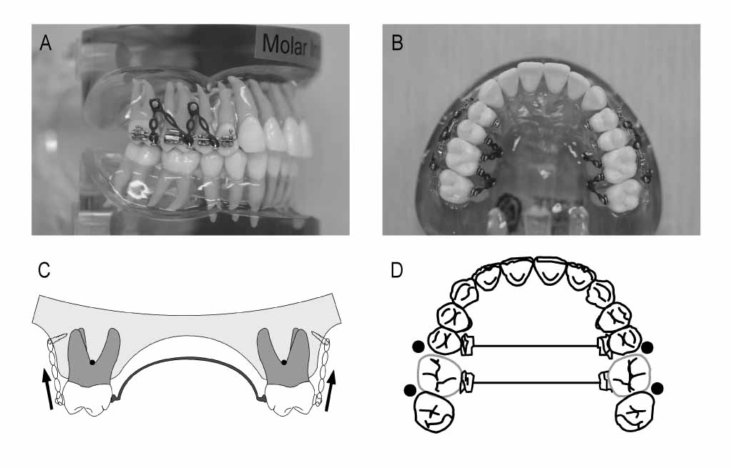
Fig 1 Appliance for intrusion of maxillary molars using miniscrew. A, Miniscrew insertion on buccal and palatal sides (buccal view); B, miniscrew insertion on buccal and palatal sides (palatal view); C, miniscrew insertion on the buccal side only, with a palatal rigid splint for preventing buccal tipping (transverse view); D, miniscrew insertion on the buccal side only, with a palatal rigid splint for preventing buccal tipping (palatal view).
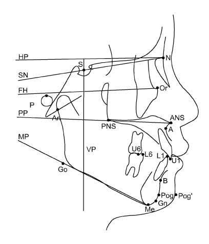
Fig 2 Cephalometric landmarks and reference planes. S (Sella), N (nasion), Ar (articulare), P (porion), Go (gonion), Me (menton), Gn (gnathion), Po (pogonion), Or (orbitale), ANS (anterior nasal spine), PNS (posterior nasal spine), A (A point), B (B point), U1 (upper incisor tip), U6 (upper molar mesiobuccal cusp tip), L1 (lower incisor tip), L6 (lower molar mesiobuccal cusp tip), Pog' (soft tissue pogonion); SN (SN plane), a line through sella and nasion; FH (FH plane), a line through orbitale and porion; HP (horizontal reference plane), a line through nasion rotated 7° from sella-nasion line; VP (vertical reference plane), a line through sella and perpendicular line to HP; PP (palatal plane), a line through ANS and PNS; MP (mandibular plane), a line through menton and gonion.
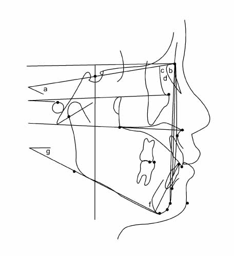
Fig 3 Skeletal angular measurements. a, SN-GoMe; b, S-N-Pog; c, SNA, S-N-A angle; d, SNB, S-N-B angle; e, ANB difference, c-d; f, IMPA, mandibular incisor long axis to Mn. plane; g, FMA, FH-MP.

Fig 4 Skeletal and dental linear measurements. 1, N-Me, Anterior facial height; 2, VP-Pog, Antero-posterior location of pogonion; 3, U6-PP (mm), perpendicular distance between mesiobuccal cusp of Mx. first molar to palatal plane; 4, U1-PP (mm), perpendicular distance between Mx. incisor edge to palatal plane; 5, OB (mm), distance between incisal edges of maxillary and mandibular central incisors, perpendicular to horizontal reference plane; 6, OJ (mm), distance between incisal edges of maxillary and mandibular central incisors, perpendicular to vertical reference plane.
Cited by 10 articles
-
Anterior open bite with temporomandibular disorders treated with intermaxillary traction using skeletal anchorage system
Hye-Sun Kim, Sang-Hoon Lee, Taegyun Youn, Hyung-Gon Kim, Jong-Ki Huh
J Korean Assoc Oral Maxillofac Surg. 2012;38(5):284-294. doi: 10.5125/jkaoms.2012.38.5.284.Anatomic study of the incisive canal in relation to midpalatal placement of mini-implant
Sung-Jin Kim, Sung-Hoon Lim
Korean J Orthod. 2009;39(3):146-158. doi: 10.4041/kjod.2009.39.3.146.Treatment of skeletal Class II adult patient with vertical and transverse problems caused by nasal airway obstruction using microimplant anchorage
Jong-Moon Chae, Na-Young Chang, Jin-Hyoung Cho, Kyung-Hwa Kang, Sang-Cheol Kim
Korean J Orthod. 2009;39(4):257-272. doi: 10.4041/kjod.2009.39.4.257.Consideration of maxillary sinus bone thickness when installing miniscrews
Do-Hyun Kim, Jin-Woo Lee, Kyung-Suk Cha, Dong-Hwa Chung
Korean J Orthod. 2009;39(6):354-361. doi: 10.4041/kjod.2009.39.6.354.Non-surgical treatment and retention of open bite in adult patients with orthodontic mini-implants
Cheol-Hyun Moon, Joo-Sin Lee, Hyun-Sun Lee, Jin-Hugh Choi
Korean J Orthod. 2009;39(6):402-419. doi: 10.4041/kjod.2009.39.6.402.Finite-element investigation of the center of resistance of the maxillary dentition
Gwang-Mo Jeong, Sang-Jin Sung, Kee-Joon Lee, Youn-Sic Chun, Sung-Seo Mo
Korean J Orthod. 2009;39(2):83-94. doi: 10.4041/kjod.2009.39.2.83.Orthodontic treatment of gummy smile by maxillary total intrusion with a midpalatal absolute anchorage system
Ryoon-Ki Hong, Seung-Min Lim, Jung-Min Heo, Seung-Hak Baek
Korean J Orthod. 2013;43(3):147-158. doi: 10.4041/kjod.2013.43.3.147.Finite element analysis of maxillary incisor displacement during en-masse retraction according to orthodontic mini-implant position
Jae-Won Song, Joong-Ki Lim, Kee-Joon Lee, Sang-Jin Sung, Youn-Sic Chun, Sung-Seo Mo
Korean J Orthod. 2016;46(4):242-252. doi: 10.4041/kjod.2016.46.4.242.Microimplant mandibular advancement (MiMA) therapy for the treatment of snoring and obstructive sleep apnea (OSA)
Joachim Ngiam, Hee-Moon Kyung
Korean J Orthod. 2010;40(2):115-126. doi: 10.4041/kjod.2010.40.2.115.Treatment of anterior open bites using non-extraction clear aligner therapy in adult patients
Heeyeon Suh, Bella Shen Garnett, Kimberly Mahood, Noor Mahjoub, Robert L. Boyd, Heesoo Oh
Korean J Orthod. 2022;52(3):210-219. doi: 10.4041/kjod21.180.
Reference
-
1. Lopez-Gavito G, Wallen TR, Little RM, Joondeph DR. Anterior open bite malocclusion: A longitudinal 10-year postretention evaluation of orthodontically treated patients. Am J Orthod. 1985. 87:175–186.
Article2. Janson G, Valarelli FP, Beltrao RT, De Freitas MR, Henriques JFC. Stability of anterior open bite extraction and nonextraction treatment in the permanent dentition. Am J Orthod Dentofacial Orthop. 2006. 129:768–774.
Article3. Janson G, Valarelli FP, Henriques JFC, De Freitas MR, Cancado RH. Stability of anterior open bite nonextraction treatment in the permanent dentition. Am J Orthod Dentofacial Orthop. 2003. 124:265–276.
Article4. Kuster R, Ingervall B. The effect of treatment of skeletal open bite with two types of bite blocks. Eur J Orthod. 1992. 14:489–499.
Article5. Rinchuse DJ. Vertical elastics for correction of anterior open bite. J Clin Orthod. 1994. 28:284.6. Chang YI, Moon SC. Cephalometric evaluation of the anterior open bite treatment. Am J Orthod Dentofacial Orthop. 1999. 115:29–38.
Article7. Denison TF, Kokich VG, Shapiro PA. Stability of maxillary surgery in openbite versus nonopenbite malocclusions. Angle Orthod. 1989. 59:5–10.8. Proffit WR, Bailey LJ, Phillips C, Turvey TA. Long-term stability of surgical open bite correction by Le Fort I osteotomy. Angle Orthod. 2000. 70:112–117.9. Ko DI, Lim SH, Kim KW. Treatment of occlusal plane canting using miniscrew anchorage. World J Orthod. 2006. 7:269–278.10. Park HS, Kwon OW, Sung JH. Nonextraction treatment of an open bite with microscrew implant anchorage. Am J Orthod Dentofacial Orthop. 2006. 130:391–402.
Article11. Park YC, Lee SY, Kim DH, Jee SH. Intrusion of posterior teeth using mini-screw implants. Am J Orthod Dentofacial Orthop. 2003. 123:690–694.
Article12. Sherwood KH, Burch JG, Thompson WJ. Closing anterior open bites by intruding molars with titanium miniplate anchorage. Am J Orthod Dentofacial Orthop. 2002. 122:593–600.
Article13. Umemori M, Sugawara J, Mitani H, Nagasaka H, Kawamura H. Skeletal anchorage system for open bite correction. Am J Orthod Dentofacial Orthop. 1999. 115:166–174.14. Xun C, Zeng X, Wang X. Microscrew anchorage in skeletal anterior open bite treatment. Angle Orthod. 2007. 77:47–56.15. Sugawara J, Baik UB, Umemori M, Takahashi I, Nagasaka H, Kawamura H, et al. Treatment and posttreatment dentoalveolar changes following intrusion of mandibular molars with application of a skeletal anchorage system (SAS) for open bite correction. Int J Adult Orthodon Orthognath Surg. 2002. 17:243–253.16. Sassouni V. A classification of skeletal facial types. Am J Orthod. 1969. 55:109–123.
Article17. Schudy FF. The rotation of the mandible resulting from growth: its implication in orthodontic treatment. Angle Orthod. 1965. 35:36–50.18. Reitan K, Rygh P. Graber TM, Vanarsdall RL, editors. Biomechanical principles and reactions. Orthodontics-current principles and techniques. 1994. 2nd ed. St. Louis: Mosby;168–169.19. Beckmann SH, Segner D. Floating norms and post-treatment overbite in open bite patients. Eur J Orthod. 2002. 24:379–390.
Article20. Proffit WR, Mason RM. Myofunctional therapy for tongue thrusting background and recommendations. J Am Dent Assoc. 1975. 90:403–411.
Article
- Full Text Links
-
- Actions
-
Cited
- CITED
-
- Close
- Share
-
- Similar articles
-
- A study on the treatment of anterior open bite with midpalatal miniscrews
- Nonextraction treatment of Class II division 2 in an adult patient using microimplant anchorage (MIA)
- Non-surgical treatment and retention of open bite in adult patients with orthodontic mini-implants
- Treatment of anterior open bite by posterior maxillary segmental osteotomy and miniplates: a case report
- Posterior open bite
