Intraspinal Clear Cell Meningioma: A Case Report
- Affiliations
-
- 1Department of Orthopedic Surgery, Yonsei University College of Medicine, Seoul, Korea. mes1007@yuhs.ac
- KMID: 1480416
- DOI: http://doi.org/10.4055/jkoa.2008.43.4.518
Abstract
- Meningioma is one of the most common tumors of the spinal canal. Spinal meningiomas are usually found in the thoracic spine and intradural extramedullary space. Intraspinal clear cell meningiomas are a rare histological variant. Fewer than 20 intraspinal cases have been reported in the literature and only two cases have been reported in the Korean literature, but there is no report available in the Korean orthopedic literature. We report here on a case of an intraspinal clear cell meningioma that was found in the thoracic region and it was completely resected. The nonspecific MR imaging characteristics make the diagnosis of this tumor difficult. Histological examination must be used to differentiate clear cell meningiomas from other tumors. Clear cell meningioma represents an aggressive variant of meningiomas, and surgical reatment and adjuvant radiotherapy are though to be essential. Further more, long term follow-up observation will be needed for detecting recurrence of clear cell meningioma.
Keyword
Figure
-
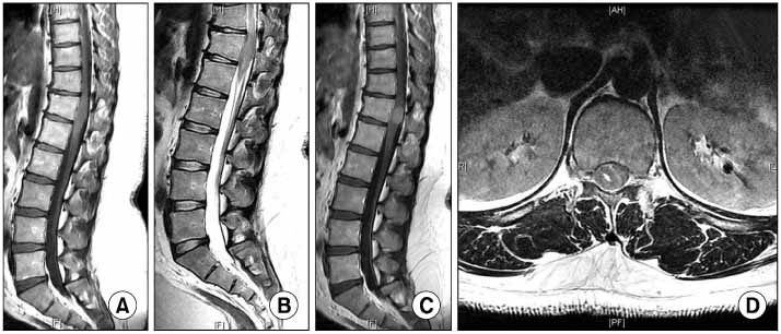
Fig. 1 (A) The preoperative T1-weighted sagittal MRI scan demonstrates that the mass shows intermediate iso-intensity signal change. About a 1.8 cm sized mass lesion is seen that involves the T12 vertebral level of the spinal canal. (B) Low signal intensity change on the T2-weighted MRI scan. (C) The T1-weighted MRI scan with gadolinium enhancement shows homogenous enhancement. (D) The mass is located in the intradural extramedullary space on an axial image.
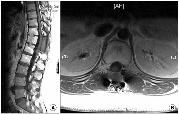
Fig. 2 (A) At the 1 year follow-up, a T1-weighted sagittal MRI scan demonstrates no evidence of tumor recurrence. (B) The T1-weighted MRI scan with gadolinium enhancement shows no residual tumor.
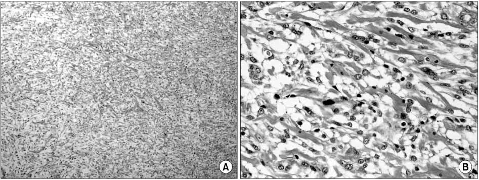
Fig. 3 (A) Photomicrograph of the spinal lesion under low magnification, demonstrates clear cells with round-to oval-shaped central nuclei and abundant clear cytoplasm, and the cells are organized in whorls and they are interspersed with hyaline sheets (Hematoxylin and Eosin; original magnification, ×100). (B) Under higher magnification, moderate unclear pleomorphism, clear cytoplasm and scatterd mitoses are seen (Hematoxylin and Eosin; original magnification, ×400).
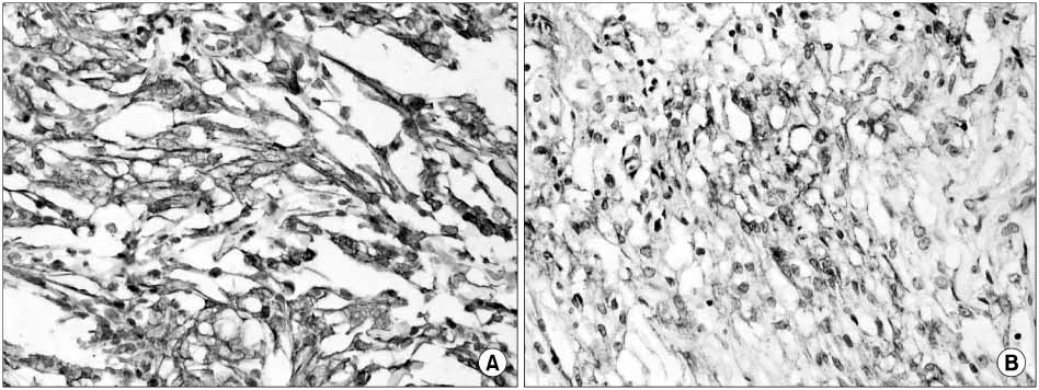
Fig. 4 (A) Strong staining is noted on the photomicrograph of the spinal lesion under vimentin immunostaining (original magnification, ×400). (B) Photomicrograph of the spinal lesion under epithelial membrane antigen immunostaining, faint membranous staining is noted (original magnification, ×400).
Reference
-
1. Cho CB, Kim JK, Cho KS, Kim DS. Clear cell meningioma of cauda equina without dural attachment. J Korean Neurosurg Soc. 2003. 34:584–585.2. Dhall SS, Tumialán LM, Brat DJ, Barrow DL. Spinal intradural clear cell meningioma following resection of a suprasellar clear cell meningioma. Case report and recommendations for management. J Neurosurg. 2005. 103:559–563.3. Epstein NE, Drexler S, Schneider J. Clear cell meningioma of the cauda equina in an adult: case report and literature review. J Spinal Disord Tech. 2005. 18:539–543.4. Jallo GI, Kothbauer KF, Silvera VM, Epstein FJ. Intraspinal clear cell meningioma: diagnosis and management: report of two cases. Neurosurgery. 2001. 48:218–221.
Article5. Kim MS, Park SH, Park YM. Thoracic intramedullary clear cell meningioma. J Korean Neurosurg Soc. 2006. 39:389–392.6. Liu PI, Liu GC, Tsai KB, Lin CL, Hsu JS. Intraspinal clear-cell meningioma: case report and review of literature. Surg Neurol. 2005. 63:285–288.
Article7. Lombardi G, Passerini A. Spinal cord tumors. Radiology. 1961. 76:381–392.
Article8. Park SH, Hwang SK, Park YM. Intramedullary clear cell meningioma. Acta Neurochir (Wien). 2006. 148:463–466.
Article9. Salvati M, Artico M, Lunardi P, Gagliardi FM. Intramedullary meningingioma: case report and review of the literature. Surg Neurol. 1992. 37:42–45.
