Distal Chevron Osteotomy for Moderate to Severe Hallux Valgus Deformity in Patients Aged 50 or Older
- Affiliations
-
- 1Department of Orthopedic Surgery, Seoul National University College of Medicine, Seoul, Korea. yoowj@snu.ac.kr
- KMID: 1480404
- DOI: http://doi.org/10.4055/jkoa.2008.43.4.445
Abstract
-
PURPOSE: The purpose of this study was to analyze the clinical and radiological results of distal chevron osteotomy in patients aged 50 and older with moderate-to-severe hallux valgus.
MATERIALS AND METHODS
The authors reviewed the medical records and radiographs of 19 patients (26 feet). Average age at time of surgery was 58 years and the mean follow-up period was 3 years and 1 month. For radiological evaluation, we analyzed changes in hallux valgus angles and 1st-2nd intermetatarsal angles after index operations. Clinical results were assessed with respect to pain, activities of daily living, and shoe-wear.
RESULTS
Hallux valgus angles and 1st-2nd intermetatarsal angles improved, but considerable correction loss occurred with time. This correction loss was found to be significantly correlated with preoperative subluxation of the 1st metatarsophalangeal joint and the 1st-2nd intermetatarsal angle. Clinically, remarkable improvements were achieved in terms of pain and level of activity, but most patients (except 4) still wore comfortable shoes rather than hard shoes at latest follow-ups.
CONCLUSION
Distal chevron osteotomy is beneficial for patients aged 50 and older with moderate-to- severe hallux valgus deformity, but correction loss may occur in patients with marked subluxation of the 1st metatarsophalangeal joint or a severe 1st-2nd intermetatarsal angle.
Keyword
MeSH Terms
Figure
-
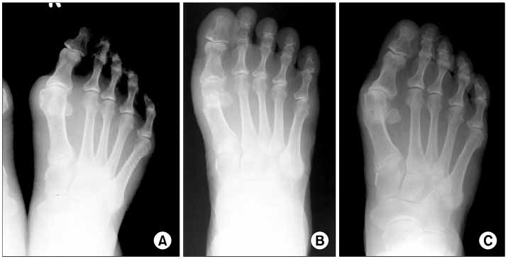
Fig. 1 A 66-year old female with hallux valgus foot deformity. (A) Preoperatively, the 1st-2nd intermetatarsal angle was 16° and 1st metatarsophalangeal joint subluxation was 12%. (B) The deformity was corrected by distal chevron osteotomy. Postoperatively, the 1st-2nd intermetatarsal angle was 4°. (C) However, 10 years after the operation, the hallux valgus angle had increased to 32°, and the 1st-2nd intermetatarsal angle to 10°, and the metatarsophalangeal joint was subluxated. Nevertheless, the patient reported only mild discomfort.
Cited by 2 articles
-
A Comparison of Proximal and Distal Chevron Osteotomy for the Correction of Severe Hallux Valgus Deformity
Hyung Seok Park, Jun Young Lee, Kang Yeol Ko, Jehong Ryu, Jae Hwan Lim
J Korean Foot Ankle Soc. 2020;24(4):129-134. doi: 10.14193/jkfas.2020.24.4.129.Comparison of Operative Results of Distal Chevron Osteotomy with and without Akin Osteotomy for Moderate to Severe Hallux Valgus
Sang Soo Park, Jun Young Lee, Woong Hee Kim
J Korean Foot Ankle Soc. 2014;18(2):56-61. doi: 10.14193/jkfas.2014.18.2.56.
Reference
-
1. Austin DW, Leventen EO. A new osteotomy for hallux valgus: a horizontally directed "V" displacement osteotomy of the metatarsal head for hallux valgus and primus varus. Clin Orthop Relat Res. 1981. 157:25–30.2. Caminear DS, Pavlovich R Jr, Pietrzak WS. Fixation of the chevron osteotomy with an absorbable copolymer pin for treatment of hallux valgus deformity. J Foot Ankle Surg. 2005. 44:203–210.
Article3. Corless JR. A modification of the mitchell procedure. J Bone Joint Surg Br. 1976. 58:138.4. Gill LH. Distal osteotomy for bunionectomy and hallux valgus correction. Foot Ankle Clin. 2001. 6:433–453.
Article5. Gill LH, Martin DF, Coumas JM, Kiebzak GM. Fixation with bioabsorbable pins in chevron bunionectomy. J Bone Joint Surg Am. 1997. 79:1510–1518.
Article6. Hattrup SJ, Johnson KA. Chevron osteotomy: analysis of factors in patient's dissatisfaction. Foot Ankle. 1985. 5:327–332.
Article7. Hirvensalo E, Böstman O, Törmälä P, Vainionpää S, Rokkanen P. Chevron osteotomy fixed with absorbable polyglycolide pins. Foot Ankle. 1991. 11:212–218.
Article8. Jahss MH, Troy AI, Kummer F. Roentgenographic and mathematical analysis of first metatarsal osteotomies for metatarsus primus varus: a comparative study. Foot Ankle. 1985. 5:280–321.
Article9. Johnson KA, Cofield RH, Morrey BF. Chevron osteotomy for hallux valgus. Clin Orthop Relat Res. 1979. 142:44–47.
Article10. Jones KJ, Feiwell LA, Freedman EL, Cracchiolo A 3rd. The effect of chevron osteotomy with lateral capsular release on the blood supply to the first metatarsal head. J Bone Joint Surg Am. 1995. 77:197–204.
Article11. Mann RA, Rudicel S, Graves SC. Repair of hallux valgus with a distal soft-tissue procedure and proximal metatarsal osteotomy. A long-term follow-up. J Bone Joint Surg Am. 1992. 74:124–129.
Article12. Mitchell LA, Baxter DE. A Chevron-Akin double osteotomy for correction of hallux valgus. Foot Ankle. 1991. 12:7–14.
Article13. Mitchell CL, Fleming JL, Allen R, Glenney C, Sanford GA. Osteotomy-bunionectomy for hallux valgus. J Bone Joint Surg Am. 1958. 40:41–58.
Article14. Peterson DA, Zilberfarb JL, Greene MA, Colgrove RC. Avascular necrosis of the first metatarsal head: incidence in distal osteotomy combined with lateral soft tissue release. Foot Ankle Int. 1994. 15:59–63.
Article15. Pochatko DJ, Schlehr FJ, Murphey MD, Hamilton JJ. Distal chevron osteotomy with lateral release for treatment of hallux valgus deformity. Foot Ankle Int. 1994. 15:457–461.
Article16. Schneider W, Aigner N, Pinggera O, Knahr K. Chevron osteotomy in hallux valgus. Ten-year results of 112 cases. J Bone Joint Surg Br. 2004. 86:1016–1020.17. Schneider W, Knahr K. Keller procedure and chevron osteotomy in hallux valgus: five-year results of different surgical philosophies in comparable collectives. Foot Ankle Int. 2002. 23:321–329.
Article18. Trnka HJ, Zembsch A, Wiesauer H, Hungerford M, Salzer M, Ritschl P. Modified Austin procedure for correction of hallux valgus. Foot Ankle Int. 1997. 18:119–127.
Article
- Full Text Links
-
- Actions
-
Cited
- CITED
-
- Close
- Share
-
- Similar articles
-
- Chevron Osteotomy as the Treatment of Moderate to Severe Hallux Valgus Deformity
- Comparison of Proximal and Modified Distal Chevron Osteotomy for the Treatment of Moderate to Severe Hallux Valgus Deformity
- Comparison of Operative Results of Distal Chevron Osteotomy with and without Akin Osteotomy for Moderate to Severe Hallux Valgus
- Comparison of Proximal Metatarsal Osteotomy andDistal Chevron Osteotomy for Correction of Hallux Valgus
- A Clinical Study of Chevron Osteotomy in Bunion - Hallux Valgus
