Inferior Sinus Venosus Type Atrial Septal Defect Initially Presenting Pulmonary Hypertension on Transthoracic Echocardiography
- Affiliations
-
- 1Division of Cardiology, Department of Internal Medicine, Gachon University Gil Hospital, Incheon, Korea. heart@gilhospital.com
- KMID: 1473767
- DOI: http://doi.org/10.4250/jcu.2009.17.1.25
Abstract
- Inferior sinus venosus type atrial septal defect (ASD) is a rare congenital cardiac deformity that occurs between the inferior vena cava and right atrium. Inferior sinus venosus defect is difficult to diagnose through transthoracic echocardiography because of its location which is infero-posterior to the fossa ovalis. Increasing pulmonary arterial pressure and pulmonary vascular resistance in patients with sinus venosus defect usually occur earlier than other types of ASD. We report a case of 19-year-old man who presented exertional dyspnea due to inferior sinus venous type ASD with mild pulmonary hypertension. In this case, we found clues from slight diastolic flattening of interventricular septum and shortened acceleration time of right ventricular outflow tract on initial transthoracic echocardiography, leading right heart catheterization and transesophageal echocardiography to reveal this rare type of ASD.
MeSH Terms
Figure
-
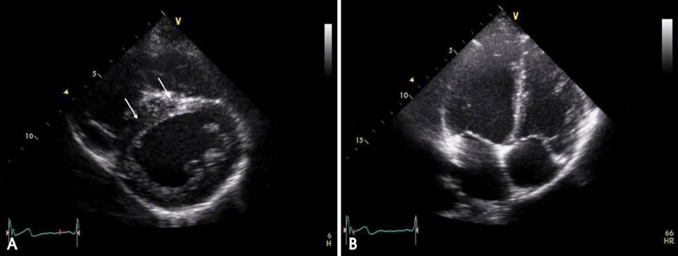
Fig. 1 2D views of transthoracic echocardiography. A: Parasternal short axis view at mid-LV level showed slight diastolic flattening of interventricular septum (arrows). B: Apical 4 chamber view showed mildly dilated right ventricle and atrium. LV: left ventricle.
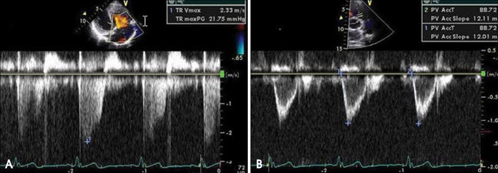
Fig. 2 Doppler findings. A: PAsP from TRV was within normal range. B: RVOT AT was 88.7 msec and calculated mPAP from the simplified Mahan's equation (mPAP= 80-[RVOT AT×0.5]) was 35.6 mmHg (B). PAsP: pulmonary arterial systolic pressure, TRV: tricuspid regurgitation velocity, RVOT: right ventricular outflow tract, AT: acceleration time, mPAP: mean pulmonary arterial pressure.
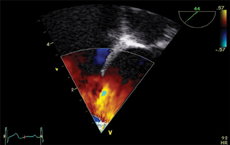
Fig. 3 Transesophageal echocardiography. Color Doppler imaging showed a defect at the extremely posterior portion of interatrial septum with left to right shunt flow.

Fig. 4 Patch repair of inferior sinus venosus type atrial septal defect (ASD) with autopericardium. The defect was located in inferior vena cava area and size was 1.5 cm. CS: coronary sinus, IVC: inferior vena cava.
Reference
-
1. Arnheid KS, Andre L, Rene P, Peter B. Inferior sinus venosus defect associated with incomplete cor triatriatum dexter and patent foramen ovale. Eur J Echocardiogr. 2006. 7:239–242.
Article2. Coon PD, Lang RM. Improved visualization of sinus venosus atrial septal defects in adults from the transthoracic approach. J Am Soc Echocardiogr. 2006. 19:1072.
Article3. Vogel M, Berger F, Kramer A, Alexi V, Lange PE. Incidence of secondary pulmonary hypertension in adults with atrial septal or sinus venosus defects. Heart. 1999. 82:30–33.
Article4. Pascoe RD, Oh JK, Warnes CA, Danielson GK, Tajik AJ, Seward JB. Diagnosis of sinus veosus atrial septal defect with transesophageal echocardiography. Circulation. 1996. 4:1049–1055.5. Oh KJ, Chung WJ, Shin MS, Han MY, Kang WC, Choi KL, Yang PS, Kim SY, Ahn TH, Shin EK. Unroofed coronary sinus associated with persistent left superior vena cava;detection by agitated saline contrast echocardiography. J Kor Soc Echocardiogr. 2004. 12:49–53.
Article
- Full Text Links
-
- Actions
-
Cited
- CITED
-
- Close
- Share
-
- Similar articles
-
- Reoperation for the Missed Inferior Sinus Venous Atrial Septal Defect
- A Case of Atrial Septal Aneurysm Associated with Atrial Septal Defect
- Role of Echocardiography in Sinus Venosus Atrial Septal Defect Combined with Systemic and Pulmonary Vascular Disease
- Surgical Repair of Inferior Sinus Venosus Defect: A Report Four Cases
- Visualization of Ostium Secundum Atrial Septal Defect by Transesophageal Echocardiography
