Right Ventricular Wall Hematoma Secondary to Percutaneous Coronary Intervention
- Affiliations
-
- 1Cardiovascular Center, Pusan National University Yangsan Hospital, Yangsan, Korea. ptca82@hotmail.com
- 2Cardiovascular Center, HanSeo Hospital, Busan, Korea.
- KMID: 1473734
- DOI: http://doi.org/10.4250/jcu.2009.17.3.99
Abstract
- Intramyocardial hematoma is known to be associated with myocardial infarction, chest trauma, coronary artery bypass operation, and complication of percutaneous coronary intervention (PCI). We describe here a rare case of 50-year-old man with a huge right ventricular (RV) wall hematoma which was newly developed two hours after PCI. The patient was treated conservatively with a successful outcome. We discuss plausible mechanisms for the development of RV wall hematoma and treatment options for the case.
MeSH Terms
Figure
-

Fig. 1 Final angiogram after stenting shows acceptable results at posterior descending artery and posterolateral branch of right coronary artery (A). Emergent coronary angiogram reveals a large amount of frank streaming of contrast material (arrows) in RV wall (B). RV: right ventricular.
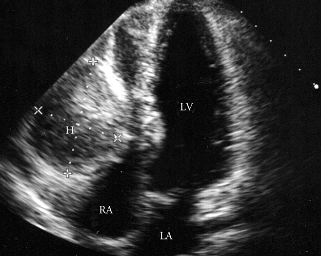
Fig. 2 Transthoracic echocardiogram from apical 4-chamber view shows a large right ventricular wall mass that almost completely obliterates right ventricular chamber. A small pericardial effusion is also present. H: hematoma, LA: left atrium, LV: left ventricle, RA: right atrium.
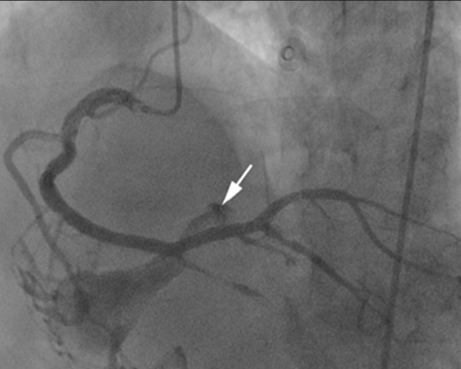
Fig. 3 Follow-up coronary angiogram shows a direct communication between hematoma and right ventricular cavity through fistula (arrow).
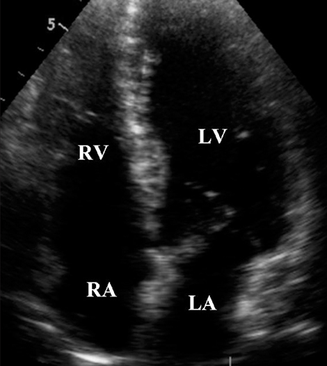
Fig. 4 A final echocardiogram at eight weeks after percutaneous coronary intervention shows a complete resolution of the hematoma. LA: left atrium, LV: left ventricle, RA: right atrium, RV: right ventricle.
Reference
-
1. Lin TH, Wu DK, Su HM, Chu CS, Voon WC, Lai WT, Sheu SH. Septum hematoma: a complication of retrograde wiring in chronic total occlusion. Int J Cardiol. 2006. 113:e64–e66.
Article2. Kawase Y, Hayase M, Ito S, Ojio S, Tahara H, Ehara M, Kondo H, Ito Y, Suzuki Y, Ishihara Y, Suzuki T. Compression of right ventricular out-flow due to localized hematoma after coronary perforation during PCI. Catheter Cardiovasc Interv. 2003. 58:202–206.
Article3. Steinwender C, Hofmann R, Leisch F. Pseudo-pericardial tamponade after perforation of the right coronary artery. Heart. 2004. 90:e36.
Article4. Rahman N, Sharif H, Jafary FH. Intramyocardial hematoma after coronary perforation during percutaneous coronary intervention--anticipated and treated. J Invasive Cardiol. 2008. 20:e224–e228.5. Drobinski G, Montalescot G, Nivet M. Intra-myocardial haemorrhage following recanalisation of a venous coronary arterial bypass by balloon angioplasty. Int J Cardiol. 1991. 31:256–258.
Article6. Cheng HW, Hung KC, Lin FC, Wu D. Spontaneous intramyocardial hematoma mimicking a cardiac tumor of the right ventricle. J Am Soc Echocardiogr. 2004. 17:394–396.
Article7. Oh DH, Doh JH, Lee SH, Kwon SU, Namgung J, Lee SY, Kim CY, Chang WI, Chang SH, Lee WR. A case of spontaneous left atrial intramural hematoma causing vaso-occlusive cardiogenic shock. J Cardiovascular Ultrasound. 2006. 14:112–115.
Article
- Full Text Links
-
- Actions
-
Cited
- CITED
-
- Close
- Share
-
- Similar articles
-
- Abdominal Wall Hematoma as a Rare Complication following Percutaneous Coronary Intervention
- A Case of Right Coronary Artery Hematoma Detected by Echocardiography after Percutaneous Coronary Intervention
- The Effect of High-Dose Valsartan on Left Ventricular Function Following Primary Percutaneous Coronary Intervention
- A Case of Extrinsic Compression of the Left Main Coronary Artery Secondary to Pulmonary Artery Dilatation
- Recent Advances in Percutaneous Coronary Intervention in Coronary Artery Disease
