A Rare Ileal Intussusception Caused by a Lipoma of the Ileum
- Affiliations
-
- 1Department of Surgery, Gumi CHA Hospital, Pochon CHA University College of Medicine, Gumi, Korea. kee39surgeon@sendu.com
- 2Department of Diagnostic Radiology, Gumi CHA Hospital, Pochon CHA University College of Medicine, Gumi, Korea.
- KMID: 1464944
- DOI: http://doi.org/10.4174/jkss.2009.77.1.59
Abstract
- Adult intussusception is a rare disease and it differs from childhood intussusception in its presentation, cause and treatment. Most of the cases have an underlying lesion within the intussusception that requires surgical resection. Making the diagnosis can be delayed because of the nonspecific and chronic symptoms, and many cases are diagnosed during performance of emergency laparotomy for treating the obstructive symptoms. A computed tomography (CT) scan is most useful for making the diagnosis of adult intussusception and is helpful in revealing the underlying lesion, although a barium enema can help to diagnose colonic intussusceptions. Surgical resection remains the recommended treatment for nearly all cases, but there is controversy about whether or not the intussusception should be initially reduced before resection. Gastrointestinal lipomas are rare benign tumors that can occur anywhere along the gut, and the small bowel is the second most common site for gastrointestinal lipomas after the colon. Intussusception of the ileum by a lipoma is very rare. We report here on a case of ileo-ileal intussusception that was caused by a lipoma of the ileum in a 35-year-old man who complained of abdominal pain of one week duration. The diagnosis of an ileo-ileal intussusception caused by a lipoma of the ileum was suspected preoperatively according to the typical CT findings, so we tried to initially reduce the intussusception during laparotomy. But manual reduction was impossible due to the edema of the lesion, and an ileum of some length had to be resected.
Keyword
MeSH Terms
Figure
-
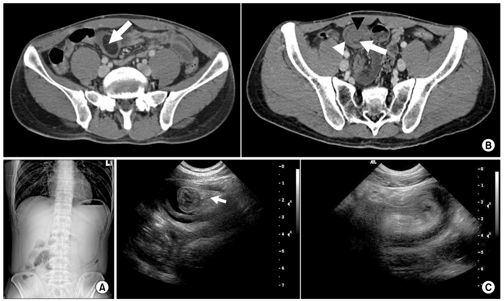
Fig. 1 (A) The plain abdominal films show the localized ileus in the lower abdomen. (B) The contrast-enhanced CT scan demonstrates an ileo-ileal intussusception caused by a mass of the ileum. (Left) The CT scan shows a homogeneous intaluminal mass with fat attenuation in the ileum (arrow). (Right) The CT scan shows a target-like appearance. The intussusceptum (black arrowhead), with an accompanying complex of mesenteric fat and blood vessels (arrow), is surrounded by the thick-walled intussuscipiens (white arrowhead). (C) Ultrasonography shows the target sign and an echogenic intraluminal mass (arrow) on the axial scan, and a sausage-shaped lesion on the longitudinal scan.
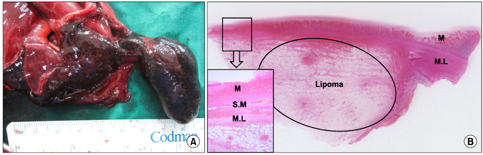
Fig. 2 Gross and microscopic findings of the specimen. (A) Photograph of the resected ileum shows a pedunculated polypoid mass, measuring 4×3×2 cm, as the lead point of the intussusception. The mass had a soft to firm, smooth surface, and the surrounding ileum and the overlying mucosa of the mass were infarcted. (B) Microscopically, a well-defined adipose mass was noted at the muscular layer and the mucosa was infarcted (H&E, ×10). Inset shows the magnified view of the lesion (H&E, ×40). M = mucosa; S.M = submucosa; M.L = muscular layer.
Reference
-
1. Azar T, Berger DL. Adult intussusception. Ann Surg. 1997. 226:134–138.2. Barussaud M, Regenet N, Briennon X, de Kerviler B, Pessaux P, Kohneh-Sharhi N, et al. Clinical spectrum and surgical approach of adult intussusceptions: a multicentric study. Int J Colorectal Dis. 2006. 21:834–839.3. Toso C, Erne M, Lenzlinger PM, Schmid JF, Buchel H, Melcher G, et al. Intussusception as a cause of bowel obstruction in adults. Swiss Med Wkly. 2005. 135:87–90.4. Kim YH, Blake MA, Harisinghani MG, Archer-Arroyo K, Hahn PF, Pitman MB, et al. Adult intestinal intussusception: CT appearances and identification of a causative lead point. Radiographics. 2006. 26:733–744.5. Kim DH, Chae GB, Choi WJ, Song TJ, Choi SY, Moon HY. Diagnosis and management of adult intussusception. J Korean Surg Soc. 1998. 55:696–704.6. Nagorney DM, Sarr MG, McIlrath DC. Surgical management of intussusception in the adult. Ann Surg. 1981. 193:230–236.7. Croome KP, Colquhoun PH. Intussusception in adults. Can J Surg. 2007. 50:E13–E14.8. Manouras A, Lagoudianakis EE, Dardamanis D, Tsekouras DK, Markogiannakis H, Genetzakis M, et al. Lipoma induced jejunojejunal intussusception. World J Gastroenterol. 2007. 13:3641–3644.9. Thompson WM. Imaging and findings of lipomas of the gastrointestinal tract. AJR Am J Roentgenol. 2005. 184:1163–1171.10. Taylor AJ, Stewart ET, Dodds WJ. Gastrointestinal lipomas: a radiologic and pathologic review. AJR Am J Roentgenol. 1990. 155:1205–1210.11. Akagi I, Miyashita M, Hashimoto M, Makino H, Nomura T, Tajiri T. Adult intussusception caused by an intestinal lipoma: report of a case. J Nippon Med Sch. 2008. 75:166–170.12. Park KT, Kim SH, Song TJ, Moon HY. Laparoscopic-assisted resection of ileal lipoma causing ileo-ileo-colic intussusception. J Korean Med Sci. 2001. 16:119–122.13. Tsushimi T, Matsui N, Kurazumi H, Takemoto Y, Oka K, Seyama A, et al. Laparoscopic resection of an ileal lipoma: report of a case. Surg Today. 2006. 36:1007–1011.14. Lin MW, Chen KH, Lin HF, Chen HA, Wu JM, Huang SH. Laparoscopy-assisted resection of ileoileal intussusception caused by intestinal lipoma. J Laparoendosc Adv Surg Tech A. 2007. 17:789–792.
- Full Text Links
-
- Actions
-
Cited
- CITED
-
- Close
- Share
-
- Similar articles
-
- Laparoscopic-assisted resection of ileal lipoma causing ileo-ileo-colic intussusception
- Endoscopic Treatment of a Symptomatic Ileal Lipoma with Recurrent Ileocolic Intussusceptions by Using Cap-Assisted Colonoscopy
- A Case of Lipoma of Terminal Ileum Causing Intussusception of the Transverse Colon
- A Case of Small Bowel Lipoma Resulting Intussusception
- Diagnosis and Treatment of Adult Intussusception Due to Gastrointestinal Lipoma
