Splenic Cavernous Lymphangioma Mimicking Splenic Hemangioma
- Affiliations
-
- 1Department of Surgery, Chonnam National University Medical School, Hwasun, Korea. koh88@dreamwiz.com
- KMID: 1464825
- DOI: http://doi.org/10.4174/jkss.2009.77.6.434
Abstract
- Lymphangioma of the spleen is a rare benign neoplasm with clinical manifestations ranging from insignificant incidental findings to large, symptomatic cystic masses requiring surgical intervention. We report a case of splenic cavernous lymphangioma mimicking splenic hemangioma. A 59-year-old woman presented with left upper quadrant pain and epigastric discomfort. Computed tomography showed a 9.5x8 cm high attenuated mass with relatively homogenous enhancement in the spleen. The initial impression was a splenic hemangioma. The patient underwent splenectomy. Gross pathologic examination revealed a 9.5x6.8x9 cm-sized fairly well circumscribed soft mass. Histologically, the tumor was composed of dilated lymphatic vessels, which contained homogenous eosinophilic material. The Final diagnosis was cavernous lymphangioma of the spleen. Herein, we report a case of splenic cavernous lymphangioma mimicking splenic hemangioma and also review the existing literature.
Keyword
MeSH Terms
Figure
-
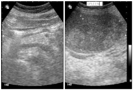
Fig. 1 Abdominal ultrasonography reveals a mild splenomegaly and about 9.2×6.6 cm-sized mixed echoic mass in the spleen.
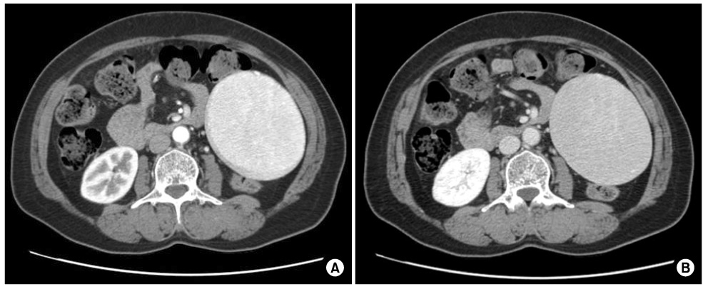
Fig. 2 The abdominal computed tomography (CT) findings. (A) In the arterial phase, CT scan shows a 9.5×8 cm high attenuated mass with relatively homogenous enhancement in the spleen. (B) In the portal pahse, CT scan shows a low attenuated mass with homogenous enhancement of similar degree to normal spleen parenchyma.
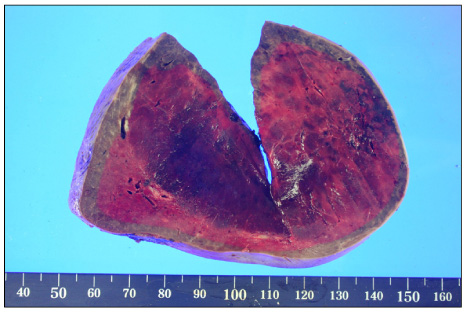
Fig. 3 Gross findings of splenic cavernous lymphangioma. Gross pathologic examination reveals a 9.5×6.8×9 cm-sized mass composed of various-sized cysts with internal hemorrhage.
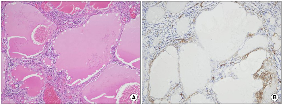
Fig. 4 Microscopic findings. (A) Histologically, the tumor is composed of dilated lymphatic vessels, and contained homogenous eosinophilic material (H&E stain, ×100). (B) Immunohistochemical finding shows positive endothelial-lined cells in immunostaining by CD 34 (×100).
Cited by 1 articles
-
A Case of Cavernous Lymphangioma of the Small Bowel Mesentery
In Taik Hong, Jae Myung Cha, Joung Il Lee, Kwang Ro Joo, Il Hyun Baek, Hyun Phil Shin, Jung Won Jeon, Jun Uk Lim
Korean J Gastroenterol. 2015;66(3):172-175. doi: 10.4166/kjg.2015.66.3.172.
Reference
-
1. Vezzoli M, Ottini E, Montagna M, La Fianza A, Paulli M, Rosso R, et al. Lymphangioma of the spleen in an elderly patient. Haematologica. 2000. 85:314–317.2. Morgenstern L, Bello JM, Fisher BL, Verham RP. The clinical spectrum of lymphangiomas and lymphangiomatosis of the spleen. Am Surg. 1992. 58:599–604.3. Komatsuda T, Ishida H, Konno K, Hamashima Y, Naganuma H, Sato M, et al. Splenic lymphangioma: US and CT diagnosis and clinical manifestations. Abdom Imaging. 1999. 24:414–417.4. Ferrozzi F, Bova D, Draghi F, Garlaschi G. CT findings in primary vascular tumors of the spleen. AJR Am J Roentgenol. 1996. 166:1097–1101.5. Hwang JK, Kim KH, Seo HJ, Kim JI, Kim JS, Yoo SJ, et al. Splenic lymphangioma of the spleen in an elderly patient. J Korean Surg Soc. 2005. 68:74–77.6. Chung JH, Suh YL, Park IA, Jang JJ, Chi JG, Kim YI, et al. A pathologic study of abdominal lymphangiomas. J Korean Med Sci. 1999. 14:257–262.7. Gorg C, Gorg K, Bert T, Barth P. Colour Doppler ultrasound patterns and clinical follow-up of incidentally found hypoechoic, vascular tumours of the spleen: evidence for a benign tumour. Br J Radiol. 2006. 79:319–325.8. Abbott RM, Levy AD, Aguilera NS, Gorospe L, Thompson WM. From the archives of the AFIP: primary vascular neoplasms of the spleen: radiologic-pathologic correlation. Radiographics. 2004. 24:1137–1163.9. Kim HS, Kim DH, Cho TY. Two cases of laparoscopic splenectomy for splenic lymphangioma. J Korean Soc Endosc Laparosc Surg. 2007. 10:118–121.
- Full Text Links
-
- Actions
-
Cited
- CITED
-
- Close
- Share
-
- Similar articles
-
- Extrinsic Indentation at Gastric Fundus by Splenic Lymphangioma
- Fine-Needle Aspiration of Splenic Lesions
- Diagnostic Usefulness of a Multimodality Study in a Mediastinal Hemangioma: A Case Report
- A Case of Splenic Pseudocyst Difficult to Differentiate from Splenic Lymphangioma
- Perirenal Lymphangioma Combined With Multiple Splenic and Hepatic Cysts
