J Korean Surg Soc.
2009 Dec;77(6):371-377. 10.4174/jkss.2009.77.6.371.
Cox-2 Expression in Malignant Breast Tumors
- Affiliations
-
- 1Department of Surgery, Paik Hospital, Inje University College of Medicine, Busan, Korea. hnkim80@hotmail.com
- 2Department of Pathology, Paik Hospital, Inje University College of Medicine, Busan, Korea.
- KMID: 1464815
- DOI: http://doi.org/10.4174/jkss.2009.77.6.371
Abstract
- PURPOSE
Cox-2 expression in breast carcinoma has been reported to be related to angiogenesis, lymph node metastasis and Her-2 expression. The aim of this study is to evaluate the clinicopathologic significance of Cox-2 expression in the invasive ductal carcinomas (IDC) and intraductal carcinomas (DCIS) of the breast.
METHODS
The materials were 353 IDC cases and 82 DCIS cases. Immunohistochemical stain for Cox-2 was interpreted as 1+ (weak & focal) and 2+ (diffuse), and the relationships between Cox-2 and ER, PR, Her-2, p53, Ki-67 and bcl-2 expressions were analyzed.
RESULTS
There was no significant difference of Cox-2 expression between IDC (148/353, 41.9%) and DCIS (38/82, 46.3%). Cox-2 (2+) expression was more frequent in low grade than intermediate and high grade IDC, but the difference was not significant statistically (P=0.0833), and there were no significant differences of Cox-2 expression according to age, tumor size, nuclear grade, lymph node metastasis in IDC and DCIS cases. In IDC cases, Cox-2 (1+ and 2+) expression showed positive relationships with p53 (+) and more than 10% of Ki-67 labeling index (P=0.0029, P=0.0015), and revealed tendencies of positive relationships with ER (+) and bcl-2 (+) (P=0.0750, P=0.0776). However, no significant relationship between Cox-2 and Her-2 expressions was recognized. In DCIS cases, Cox-2 (2+) expression rate was increased in cases showing negative for Her-2 (P=0.0092) and positive for bcl-2 (P=0.0486).
CONCLUSION
Cox-2 expression seems to be involved in the development of breast carcinomas, but not related to the invasiveness. Cox-2 expression, especially 2+, in the DCIS cases suggest a possibility of less aggressive biological behavior.
Keyword
MeSH Terms
Figure
-
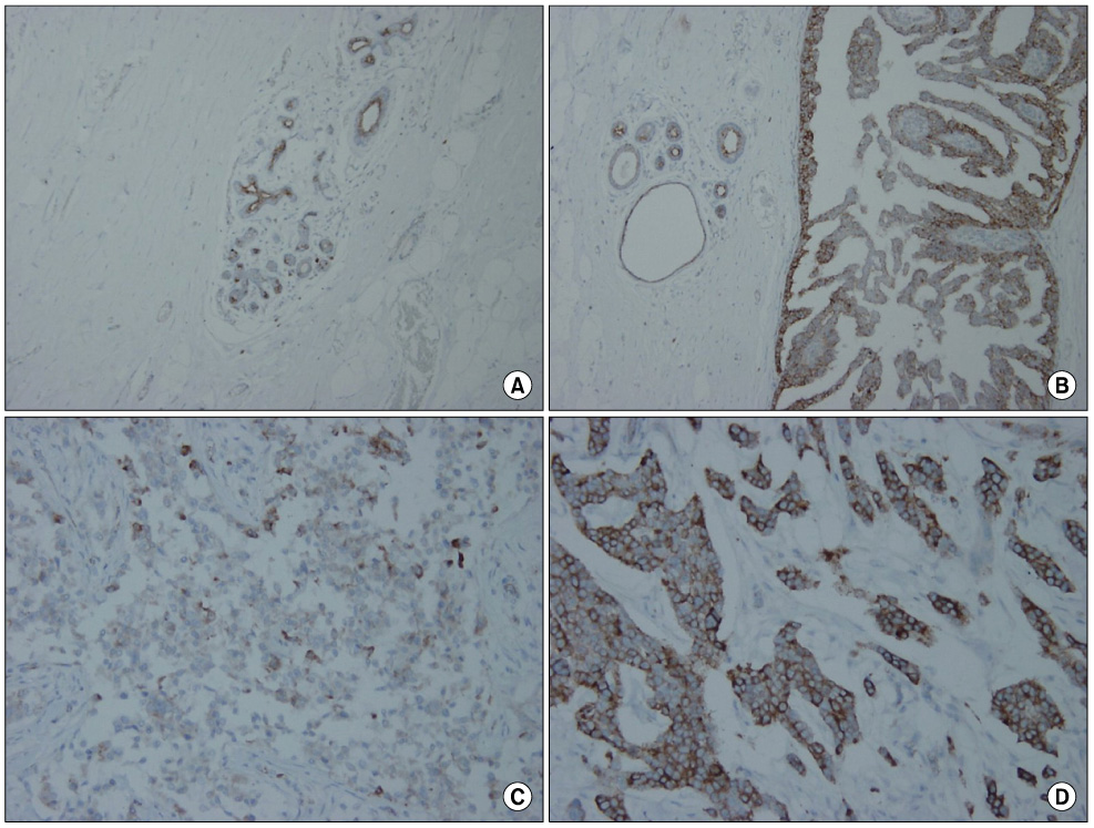
Fig. 1 Cox-2 expressions in the malignant breast lesions. Normal ductal tissue shows focal expression along the luminal borders (A, ×40). Uneven expression in ducal carcinoma in situ (B, ×100). Focal expression (C) or diffuse expression (D) in the invasive ductal carcinomas (×200).
Reference
-
1. Kelly LM, Hill AD, Kennedy S, Connolly EM, Ramanath R, Teh S, et al. Lack of prognostic effect of Cox-2 expression in primary breast cancer on short-term follow-up. Eur J Surg Oncol. 2003. 29:707–710.2. Costa C, Soares R, Reis-Filho JS, Leitao D, Amendoeira I, Schmitt FC. Cyclo-oxygenase 2 expression is associated with angiogenesis and lymph node metastasis in human breast cancer. J Clin Pathol. 2002. 55:429–434.3. Park K, Han S, Shin E, Kim HJ, Kim JY. Cox-2 expression on tissue microarray of breast cancer. Eur J Surg Oncol. 2006. 32:1093–1096.4. Wulfing P, Diallo R, Muller C, Wulfing C, Poremba C, Heinecke A, et al. Analysis of cyclooxygenase-2 expression in human breast cancer: high throughput tissue microarray analysis. J Cancer Res Clin Oncol. 2003. 129:375–382.5. Khuder SA, Mutgi AB. Breast cancer and NSAID use: a meta-analysis. Br J Cancer. 2001. 84:1188–1192.6. Witton CJ, Hawe SJ, Cooke TG, Bartlett JM. Cyclooxygenase 2 (COX2) expression is associated with poor outcome in ER-negative, but not ER-positive, breast cancer. Histopathology. 2004. 45:47–54.7. Half E, Tang XM, Gwyn K, Sahin A, Wathen K, Sinicrope FA. Cyclooxygenase-2 expression in human breast cancers and adjacent ductal carcinoma in situ. Cancer Res. 2002. 62:1676–1681.8. Liu CH, Chang SH, Narko K, Trifan OC, Wu MT, Smith E, et al. Overexpression of cyclooxygenase-2 is sufficient to induce tumorigenesis in transgenic mice. J Biol Chem. 2001. 276:18563–18569.9. Sano H, Kawahito Y, Wilder RL, Hashiramoto A, Mukai S, Asai K, et al. Expression of cyclooxygenase-1 and -2 in human colorectal cancer. Cancer Res. 1995. 55:3785–3789.10. Chan TA, Morin PJ, Vogelstein B, Kinzler KW. Mechanisms underlying nonsteroidal antiinflammatory drug-mediated apoptosis. Proc Natl Acad Sci U S A. 1998. 95:681–686.11. Sheng H, Shao J, Morrow JD, Beauchamp RD, DuBois RN. Modulation of apoptosis and Bcl-2 expression by prostaglandin E2 in human colon cancer cells. Cancer Res. 1998. 58:362–366.12. Harris RE, Robertson FM, Abou-Issa HM, Farrar WB, Brueggemeier R. Genetic induction and upregulation of cyclooxygenase (COX) and aromatase (CYP19): an extension of the dietary fat hypothesis of breast cancer. Med Hypotheses. 1999. 52:291–292.13. Gately S. The contributions of cyclooxygenase-2 to tumor angiogenesis. Cancer Metastasis Rev. 2000. 19:19–27.14. Ristimaki A, Sivula A, Lundin J, Lundin M, Salminen T, Haglund C, et al. Prognostic significance of elevated cyclooxygenase-2 expression in breast cancer. Cancer Res. 2002. 62:632–635.15. Denkert C, Winzer KJ, Muller BM, Weichert W, Pest S, Kobel M, et al. Elevated expression of cyclooxygenase-2 is a negative prognostic factor for disease free survival and overall survival in patients with breast carcinoma. Cancer. 2003. 97:2978–2987.16. Nassar A, Radhakrishnan A, Cabrero IA, Cotsonis G, Cohen C. COX-2 expression in invasive breast cancer: correlation with prognostic parameters and outcome. Appl Immunohistochem Mol Morphol. 2007. 15:255–259.17. Schreinemachers DM, Everson RB. Aspirin use and lung, colon, and breast cancer incidence in a prospective study. Epidemiology. 1994. 5:138–146.18. Harris RE, Namboodiri KK, Farrar WB. Nonsteroidal antiinflammatory drugs and breast cancer. Epidemiology. 1996. 7:203–205.19. Sharpe CR, Collet JP, McNutt M, Belzile E, Boivin JF, Hanley JA. Nested case-control study of the effects of non-steroidal anti-inflammatory drugs on breast cancer risk and stage. Br J Cancer. 2000. 83:112–120.
- Full Text Links
-
- Actions
-
Cited
- CITED
-
- Close
- Share
-
- Similar articles
-
- Significance of c-kit and COX-2 Expression in Breast Tissue
- Patterns of p53 expression in phyllodes tumors of the breast: an immunohistochemical study
- Expression of Cyclooxygenase-2 in Human Breast Carcinoma: Relevance to Tumor Angiogenesis and Expression of Estrogen Receptor
- Cyclooxygenase-2 Expression in Ovarian Tumors
- Correlation between COX-2 Expression and Hormone Receptors in Invasive Ductal Breast Cancer
