Omental Actinomycosis Coexisting with Colon Cancer
- Affiliations
-
- 1Department of Surgery, College of Medicine, Chung-Ang University, Seoul, Korea. limmhm@kornet.net
- 2Department of Pathology, College of Medicine, Chung-Ang University, Seoul, Korea.
- KMID: 1464794
- DOI: http://doi.org/10.4174/jkss.2009.77.Suppl.S17
Abstract
- Actinomycosis is a rare infection caused by Actinomyces species, normal commensal inhabitants of the human bronchial and gastrointestinal tract. Infection occurs after preceding mucosal break-down by variable causes. A preoperative diagnosis is difficult because of its nonspecific clinical features, mimicking malignancy, tuberculosis or other inflammatory diseases. We report a case of abdominal actinomycosis presenting as an omental mass, which coexists with ascending colon cancer. Actinomycosis was diagnosed by histopathologic demonstration of sulfur granules in a specimen resected by laparoscopic exploration. Following surgery, the patient was treated with IV penicillin (20 million IU/day) for 3 weeks, and follow-up colonoscopy showed adenocarcinoma in the ascending colon. The patient underwent right hemicolectomy, then treated with intravenous penicillin for 4 weeks postoperatively and oral penicillin for 6 months. The patient has been free of recurrence for 6 months.
Keyword
MeSH Terms
Figure
-
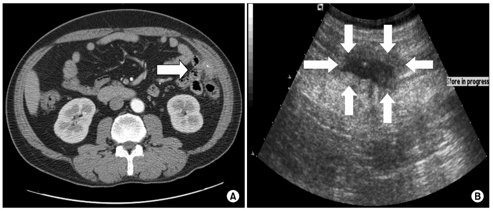
Fig. 1 (A) Abdomino-pelvic computed tomography shows 3.3×2 cm ill defined soft tissue density in left-sided omentum with enhancement and 1.7 cm internal linear high density (white arrow). (B) Abdominal ultrasonography shows about 3 cm infiltrative ill defined hypoechoic lesion in left abdominal wall and omentum (white arrows).
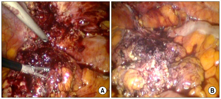
Fig. 2 (A) Omental abscess resected by laparoscopic approach. (B) Intraoperative finding shows multiple small abscess formations located through omentum.
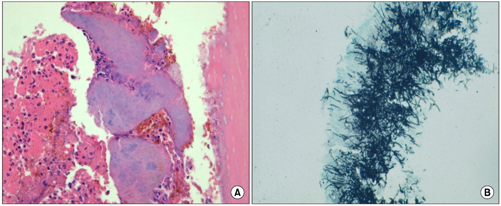
Fig. 3 Lesion shows characteristic surfur granules and inflammatory reaction (H&E, ×200) (A). Sulfur granule is composed of radiating small slender fillamentous organisms (Gomori methenamine silver stain, ×400) (B).
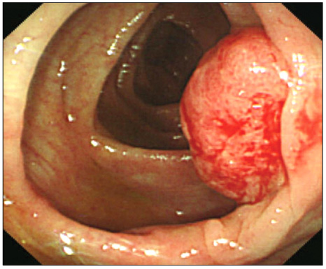
Fig. 4 Colonoscopic examination shows 2 cm protruding mass lesion in ascending colon, suggesting cancer.
Reference
-
1. Joo YT. Abdominal actinomycosis presented as a periappendiceal abscess. J Korean Surg Soc. 2004. 67:342–345.2. Kaya M, Sakarya MH. A rare cause of chronic abdominal pain, weight loss and anemia: abdominal actinomycosis. Turk J Gastroenterol. 2007. 18:254–257.3. Filippou D, Psimitis I, Zizi D, Rizos S. A rare case of ascending colon actinomycosis mimicking cancer. BMC Gastroenterol. 2005. 5:1.4. Hefny AF, Joshi S, Saadeldin YA, Fadlalla H, Abu-Zidan FM. Primary anterior abdominal wall actinomycosis. Singapore Med J. 2006. 47:419–421.5. Kim SY, Lee HS, Kim SM, Lee WJ, Lee JY, Choi SJ, et al. A case of abdominal actinomycosis presenting as mesenteric mass. Korean J Gastroenterol. 2008. 51:48–51.6. Lee SG, Roh YH, Park KJ, Choi HJ, Jung GJ, Han MS. The clinical study of abdominopelvic actinomycosis. J Korean Surg Soc. 2006. 70:47–52.7. Jung EY, Choi SN, Park DJ, You JJ, Kim HJ, Chang SH. Abdominal actinomycosis associated with a sigmoid colon perforation in a patient with a ventriculoperitoneal shunt. Yonsei Med J. 2006. 47:583–586.8. Filipovic B, Milinic N, Nikolic G, Ranthelovic T. Primary actinomycosis of the anterior abdominal wall: case report and review of the literature. J Gastroenterol Hepatol. 2005. 20:517–520.9. Karagulle E, Turan H, Turk E, Kiyici H, Yildirim E, Moray G. Abdominal actinomycosis mimicking acute appendicitis. Can J Surg. 2008. 51:E109–E110.
