Lab Anim Res.
2010 Mar;26(1):47-53. 10.5625/lar.2010.26.1.47.
Inhibitory Effect of Luteolin Liposome Solution by Animal Model for Atopic Dermatitis in NC/Nga Mice
- Affiliations
-
- 1Nabion Corporation, Seoul, Korea.
- 2Department of Chemical Engineering, Hanyang University, Seoul, Korea.
- 3Department of Applied Chemistry, Dongduk Women's University, Seoul, Korea.
- 4Department of Public Health, Keimyung University, Daegu, Korea. kim9399@kmu.ac.kr
- KMID: 1458939
- DOI: http://doi.org/10.5625/lar.2010.26.1.47
Abstract
- Atopic dermatitis is a common chronic inflammatory skin disease, associated with marked inflammatory cells (of mast cells and eosinophils) and severe itching, which leads to clinical problems in the pediatric population. This study was designed to investigate the inhibitory effects of luteolin liposome solution, that is entrapped the hydrophobic luteolin (one of the flavonoids) into ethosome to improve its stability, by using hapten-induced atopic dermatitis animal model (NC/Nga mice).The luteolin liposome treated mice showed anti-inflammatory effect as evidenced by the lowering of erythema and edema in clinical observation, reduction of inflammatory cell infiltration and epidermal thickness in histopathological examination, when compared with TNCB induced controls. Luteolin liposome solution also reduced the serum IgE level which played important roles in the atopic dermatitis model. These results suggest that luteolin liposome solution has some merit in this formulation showing inhibitory effects for the atopic dermatitis.
Keyword
MeSH Terms
Figure
-
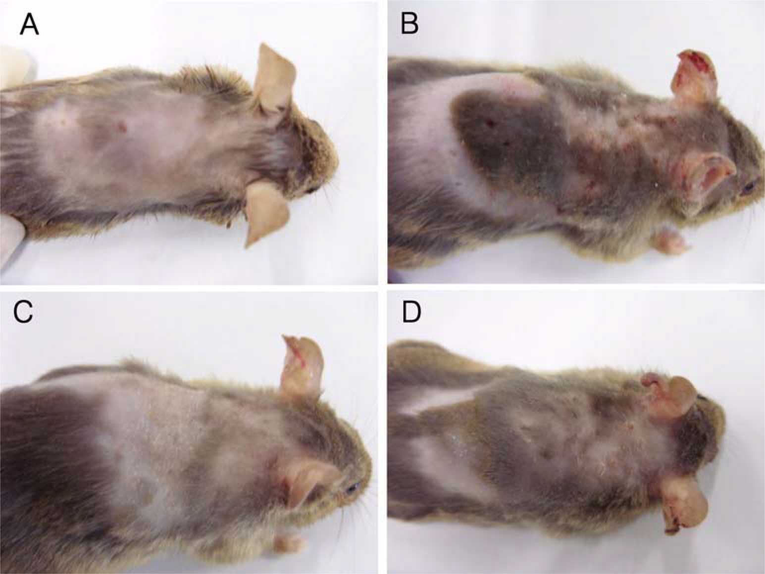
Figure 1. Gross lesions of NC/Nga mouse skin. A: Normal control, B: TNCB induced control, C: Luteolin liposome solution, D: Luteolin.
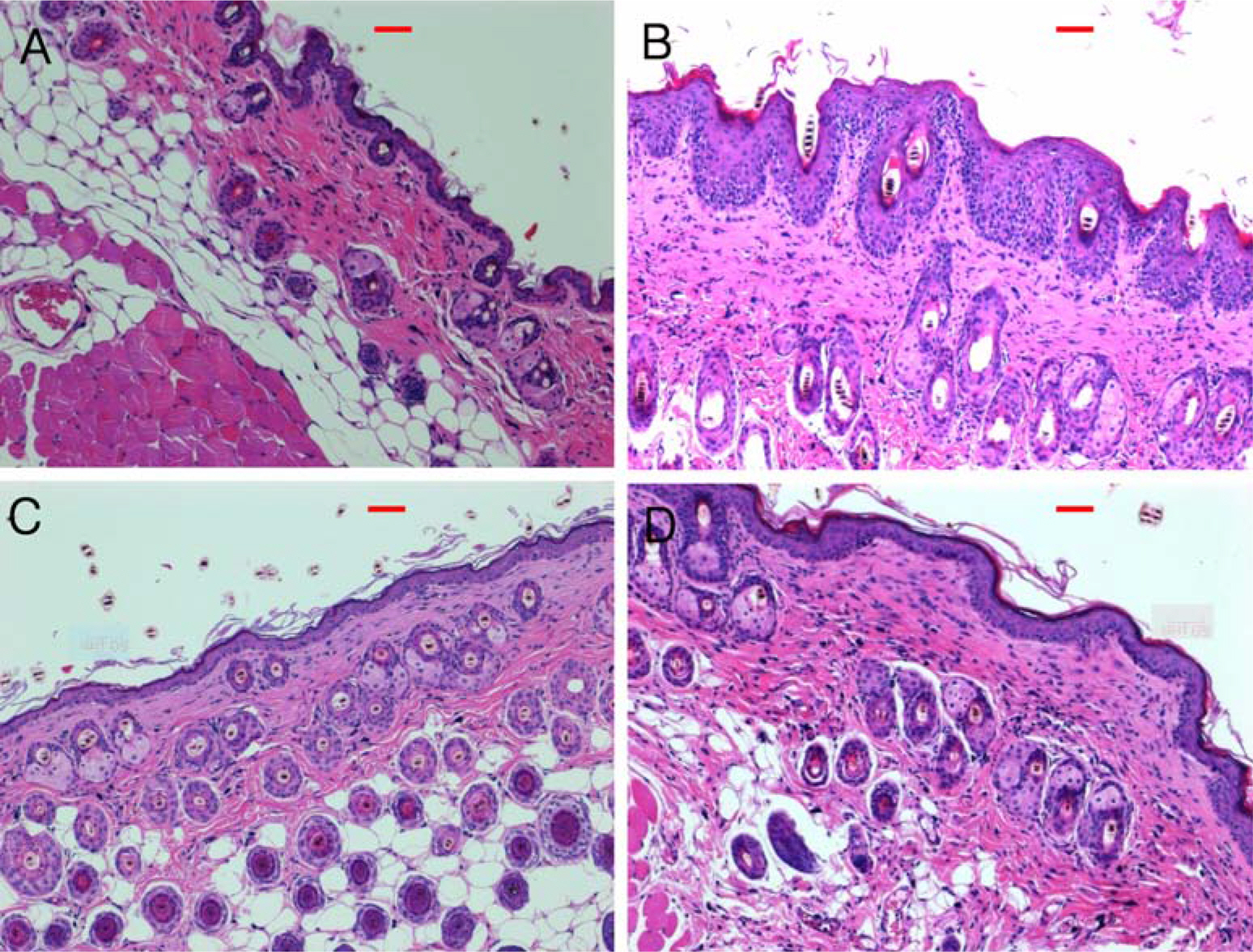
Figure 2. Histological observation of NC/Nga mouse skin by H&E staining (×100) (red bar=50 µm). A: Normal control, B: TNCB induced control, C: Luteolin liposome solution, D: Luteolin.
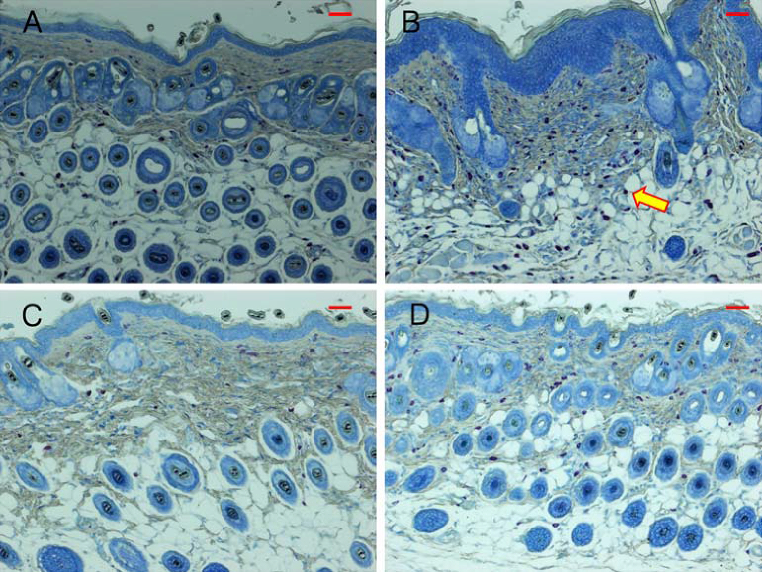
Figure 3. Histological observation of NC/Nga mouse skin by Toluidine blue staining (×100) (red bar=50 µm). A: Normal control, B: TNCB induced control, C: Luteolin liposome solution, D: Luteolin. There are many mast cells (arrow) in TNCB control (B).
Reference
-
Falus A.., Merétey K.1992. Histamine: an early messenger in inflammatory and immune reactions. Immunol. Today. 13(5):154–156.Friedman P.S.2006. Contact sensitization and allergic contact dermatitis immunobiological mechanisms. Toxicol. Lett. 162:49–54.Furue M.., Terao H.., Moroi Y.., Koga T.., Kubota Y.., Nakayama J.., Furukawa F.., Tanaka Y.., Katayama I.., Kinukawa N.., Nose Y.., Urabe K.2004. Dosage and adverse effects of topical tacrolimus and steroids in daily management of atopic dermatitis. J. Dermatol. 31(4):277–283.
ArticleGoutet M.., Pépin E.., Langonné I.., Huguet N.., Ban M.2005. Identification of contact and respiratory sensitizers using flow cytometry. Toxicol. Appl. Pharmacol. 205(3):259–270.
ArticleHruza L.L.., Pentland A.P.1993. Mechanism of UV induced inflammation. J. Invest. Dermatol. 15(2):111–115.Kaefer C.M.., Milner J.A.2008. The role of herbs and spices in cancer prevention. J. Nutri. Biochem. 19:347–361.
ArticleLee S.H.., Baek S.J.., Kim H.A.., Heo Y.2006. 2,4-dinitrochlorobenzene-induced atopic dermatitis like immune alteration in mice. J. Toxicol. Pub. Health. 22(4):357–364.Malaviya R.., Morrison A.R.., Pentland A.P.1996. Histamine in human epidermal cells is induced by ultraviolet light injury. J. Invest. Dermatol. 106(4):785–789.
ArticleMatsuda H.., Watanabe N.., Geba G.P.., Sperl J.., Tsudzuki M.., Hiroi J.., Matsumoto M.., Ushio H.., Saito S.., Askenase P.W.., Ra C.1997. Development of atopic dermatitis-like skin lesion with IgE hyperproduction in NC/Nga mice. Int. Immunol. 9(3):461–466.
ArticleMatsumoto M.., Ra C.., Kawamoto K.., Sato H.., Itakura A.., Sawada J.., Ushio H.., Suto H.., Mitsuishi K.., Hikasa Y.., Matsuda H.1999. IgE hyperproduction through enhanced tyrosine phosphorylation of Janus kinase 3 in NC/Nga mice, a model for human atopic dermatitis. J. Immunol. 162(2):1056–1063.McGirt L.Y.., Beck L.A.2006. Innate immune defects in atopic dermatitis. J. of Allergy and Clin. Immunol. 118(1):202–208.
ArticleMenzies-Gow A.., Ying S.., Sabroe I.., Stubbs V.L.., Soler D.., Williams T.J.., Kay A.B.2002. Eotaxin (CCL11) and eotaxin-2 (CCL24) induce recruitment of eosinophils, basophils, neutrophils, and macrophages as well as features of early- and late-phase allergic reactions following cutaneous injection in human atopic and nonatopic volunteers. J. Immunol. 169(5):2712–2718.
ArticleMueller D.L.., Jenkins M.K.., Schwartz R.H.1989. Clonal expansion versus functional clonal inactivation: a costimulatory signalling pathway determines the outcome of T cell antigen receptor occupancy. Annu. Rev. Immunol. 7:445–480.
ArticlePlitnick L.M.., Loveless S.E.., Ladics G.S.., Holsapple M.P.., Selgrade M.J.., Sailstad D.M.., Smialowicz R.J.2002. Cytokine profiling for chemical sensitizers: application of the ribonuclease protection assay and effect of dose. Toxicol. Appl. Pharmacol. 179(3):145–154.
ArticleRenz H.., Brodie C.., Bradley K.., Leung D.Y.., Gelfand E.W.1994. Enhancement of IgE production by anti-CD40 antibody in atopic dermatitis. J. Allergy Clin. Immunol. 93(3):658–668.
ArticleSchuller E.., Oppel T.., Bornhövd E.., Wetzel S.., Wollenberg A. 2004. Tacrolimus ointment causes inflammatory dendritic epidermal cell depletion but no Langerhans cell apoptosis in patients with atopic dermatitis. J. Allergy and Clin. Immunol. 114(1):137–143.
ArticleSchultz-Larsen F.., Hanifin J.M.2002. Epidemiology of atopic dermatitis. Immunol. Allergy Clin. North Am. 22:1–24.Shiohara T.., Hayakawa J.., Mizukawa Y.2004. Animal models for atopic dermatitis: are they relevant to human disease? J. Dermatol. Sci. 36(1):1–9.
ArticleTaniguchi Y.., Kohno K.., Inoue S.., Koya-Miyata S.., Okamoto I.., Arai N.., Iwaki K.., Ikeda M.., Kurimoto M.2003. Oral administration of royal jelly inhibits the development of atopic dermatitis-like skin lesions in NC/Nga mice.Touitou E.., Dayan N.., Bergelson L.., Godin B.., Eliaz M.2000. Ethosomes-novel vesicular carriers for enhanced delivery: characterization and skin penetration properties J. Control. Release. 65:403–418.Touitou E.., Meidan V.M.., Horwitz E.1998. Methods for quantitative determination of drug localized in the skin. J. Control. Release. 56:7–21.
ArticleWisselink M.A.., Willemse T. 2009. The efficacy of cyclosporine A in cats with presumed atopic dermatitis: A double blind, randomised prednisolone-controlled study. The Vet. Journal. 180:55–59.
ArticleYano S.., Umeda D.., Yamashita S.., Yamada K.., Tachibana H.2009. Dietary apigenin attenuates the development of atopic dermatitis-like skin lesions in NC/Nga mice. J. Nutri. Biochem. 20(11):876–881.
ArticleYing S.., Robinson D.S.., Meng Q.., Barata L.T.., McEuen A.R.., Buckley M.G.., Walls A.F.., Askenase P.W.., Kay A.B.1999. C-C chemokines in allergen-induced late-phase cutaneous responses in atopic subjects: association of eotaxin with early 6-hour eosinophils, and of eotaxin-2 and monocyte chemoattractant protein-4 with the later 24-hour tissue eosinophilia, and relationship to basophils and other C-C chemokines (monocyte chemoattractant protein-3 and RANTES). J. Immunol. 163(7):3976–3984.Yun J.W.., Kim H.., Kang H.J.., Koh J.Y.., Kim B.H.2008. Models for Atopic Dermatitis in NC/Nga Mice. Lab. Animal Research. 24(1):59–66.
- Full Text Links
-
- Actions
-
Cited
- CITED
-
- Close
- Share
-
- Similar articles
-
- A Novel Model for Human Atopic Dermatitis: Application of Repeated DNCB Patch in BALB/c Mice, in Comparison with NC/Nga Mice
- A Study on Tests of Skin Safety and Inhibition of Atopic Dermatitis Using a StoneTouch(R) Infrared Scanner in a Mouse Model
- Developing an Atopic Dermatitis Model and the Effects of Actinidia Extract on Dermatitis in NC/Nga Mice
- Alteration of Innate Immune T and B Cells in the NC/Nga Mouse
- Anti-inflammatory and Anti-pruritic Effects of Portulaca oleracea L. Extract Using In Vitro and In Vivo Inflammation Model: LPS-treated Raw264.7 Cells, Keratinocytes, NC/Nga Mice and Hairless SKH-1 Mice
